Molecular pathology research services
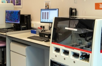
Our state-of-the-art facility offers high-throughput, rapid immunohistochemical techniques for research specimens.
Our capabilities

We provide a comprehensive digital pathology workflow—from staining to slide scanning and image analysis—using our custom-developed multiplex antibody panels in human tissues.
We have extensive experience working with a wide range of tissue types, including oesophageal, pancreatic, bladder, prostate, placental, ovarian, and endometrial samples.
Our skilled and collaborative team partners with researchers to develop multiplex panels of up to 6 markers, utilizing the Leica Bond RX and the Akoya Biosciences PhenoImager HT platforms. The lab operates in compliance with HTA regulations, employs Elab for sample tracking, and adheres to Good Laboratory Practice (GLP) standards.
Services offered

- Staining: Chromogenic and immunofluorescent staining using the automated Leica Bond RX
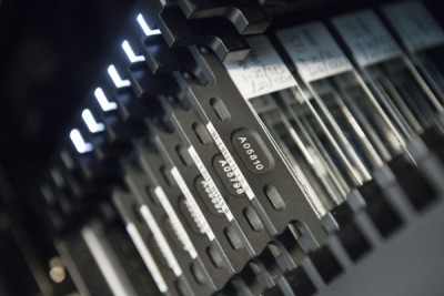
- Slide Scanning: Brightfield and fluorescence imaging with the PhenoImager HT
- Image Analysis: AI-driven analysis using inForm and QuPath.
Contact us
For more information about project timelines and pricing, please get in touch with Sunny Sunshine at S.Sunshine@surrey.ac.uk.