New computer model of lung tissue could herald safer radiotherapy for cancer
An innovative computer model of a human lung is helping scientists simulate, for the first time, how a burst of radiation interacts with the organ on a cell-by-cell level.
This research, carried out at the University of Surrey and GSI Helmholtzzentrum für Schwerionenforschung, Darmstadt, could lead to more targeted treatments for cancer and reduce the damage caused by radiotherapy.

Nowadays, more than half of cancer patients receive radiotherapy – but too high a dose can injure their lungs. This can lead to conditions like pneumonitis and fibrosis.
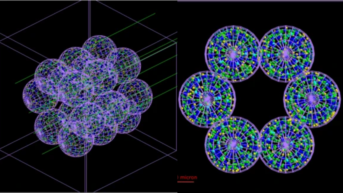
To study these injuries, researchers at GSI and the University of Surrey used artificial intelligence to develop a new model of part of a human lung – cell by cell.
For the first time, BioDynaMo makes interactive models of entire human organs achievable. This will allow us to model individual patients’ lungs in a way that’s just not possible with the very general statistical methods we currently use.Professor Dr Marco Durante, Head of the Biophysics Department, GSI Helmholtzzentrum für Schwerionenforschung
What’s more – it will allow us to study the way fibrosis and other conditions are actually caused, and how they develop over time.

The research is published in the journal Communications Medicine.
It helps to promote UN Sustainable Development Goals (SDGs) 3 (good health and well-being) and 9 (industry, innovation and infrastructure).
Media Contacts
External Communications and PR team
Phone: +44 (0)1483 684380 / 688914 / 684378
Email: mediarelations@surrey.ac.uk
Out of hours: +44 (0)7773 479911