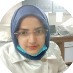
Dr Reshma Bano
About
Biography
Reshma Bano is a postdoctoral research fellow at the Department of Physics, University of Surrey. She earned her PhD in Biosciences at Jamia Millia Islamia, New Delhi, India, under the supervision of Dr Mohd Mohsin, focusing on the construction of FRET-based genetically encoded nanosensors. Reshma’s current postdoctoral research endeavours are dedicated to pioneering cutting-edge advances in the field of Nucleic Acid Nanotechnology Originated Protocells (NANOPCs). Her project work involves the encapsulation of RNA transcription machinery within liposomes functionalised with DNA-based synthetic nanopores, opening up new horizons in biotechnology and nanomedicine. Reshma's another research, committed to unravelling the complexities of heterogeneity in synthetic and living systems, by creating artificial cell.
ResearchResearch interests
My research interests lie at the intersection of biomaterials, biophysics, and membrane biology, with a particular focus on synthetic membrane systems such as Giant Unilamellar Vesicles (GUVs). I am especially interested in understanding the structural and functional dynamics of lipid membranes, their interaction with proteins and small molecules, and their application in modeling cellular processes.
Through my current project, I have gained expertise in techniques such as the inverted emulsion method for GUV preparation, fluorescence microscopy, and lipid-based assay development. My broader goal is to contribute to the development of minimal cell systems and membrane-based platforms for drug delivery, biosensing, and synthetic biology.
During my PhD, I focused on the isolation and characterization of exosomes and designed FRET-based tools for sensing biological interactions. In the future, I am also keen to explore the development and application of artificial exosomes as targeted delivery systems and diagnostic tools, combining my expertise in bioengineering and nanoscale sensing.
Looking ahead, I aim to expand this research by integrating microfluidic technologies and molecular probes to study membrane mechanics and synthetic cell communication in more physiologically relevant contexts.
Research interests
My research interests lie at the intersection of biomaterials, biophysics, and membrane biology, with a particular focus on synthetic membrane systems such as Giant Unilamellar Vesicles (GUVs). I am especially interested in understanding the structural and functional dynamics of lipid membranes, their interaction with proteins and small molecules, and their application in modeling cellular processes.
Through my current project, I have gained expertise in techniques such as the inverted emulsion method for GUV preparation, fluorescence microscopy, and lipid-based assay development. My broader goal is to contribute to the development of minimal cell systems and membrane-based platforms for drug delivery, biosensing, and synthetic biology.
During my PhD, I focused on the isolation and characterization of exosomes and designed FRET-based tools for sensing biological interactions. In the future, I am also keen to explore the development and application of artificial exosomes as targeted delivery systems and diagnostic tools, combining my expertise in bioengineering and nanoscale sensing.
Looking ahead, I aim to expand this research by integrating microfluidic technologies and molecular probes to study membrane mechanics and synthetic cell communication in more physiologically relevant contexts.
Publications
The emergence of the novel coronavirus, SARS-CoV-2, in 2019 has led to a global pandemic, affecting individuals across all age groups. As a member of the coronaviridae family, SARS-CoV-2 is a positive-sense RNA virus with helical symmetry nucleocapsid and distinctive spike proteins facilitating host cell binding. This virus shares characteristics with other coronaviruses, including Middle East respiratory syndrome (MERS-CoV) and severe acute respiratory syndrome (SARS-CoV), causing a spectrum of illnesses from mild cold-like symptoms to severe respiratory distress and organ failure. Common symptoms of COVID-19 include fever, dry cough, fatigue, and various other manifestations affecting multiple organ systems. The detection and diagnosis of COVID-19 rely on diverse methods, including nucleic acid-based testing, imaging, antigen or antibody testing, and emerging biosensing tools. These techniques enable the rapid identification of the virus in infected individuals, aiding in patient management and public health efforts. Additionally, the angiotensin-converting enzyme 2 (ACE2) receptor affinity of the spike protein contributes to viral cell entry, highlighting potential targets for therapeutic intervention. This chapter reviews current coronavirus detection methods and anticipates future advancements for early-stage virus detection. The development of innovative sensing techniques based on genomic and structural details of the virus holds promise for enhancing diagnostic capabilities and improving patient outcomes in the ongoing battle against COVID-19.
Nanovesicles, including exosomes, microvesicles, and apoptotic bodies, serve as crucial mediators of intercellular communication by transporting biological material between cells. Despite their shared impact on host immunity, nanovesicles exhibit diverse biogenesis and distinct protein compositions. Exosomes play a crucial role in cellular cargo transport and are gaining attention as potential diagnostic markers and therapeutic targets for cancer. However, isolating highly pure subpopulations of nanovesicles poses a significant challenge due to their similarity in size to other contaminants. Researchers are continuously developing innovative approaches for nanovesicle isolation and analysis to overcome these challenges. Recent literature highlights a shift toward embracing novel nanosensors for the isolation, quantification, and characterization of nanovesicles. This chapter explores advancements in fluorescence sensors, revolutionizing nanovesicle analysis by reducing sample volume requirements and enhancing isolation precision. The combination of biosensor and fluorescent technologies demonstrates unparalleled selectivity and sensitivity in detecting and quantifying extracellular vesicles (EVs), offering boundless promise in biomedicine and clinical translation. These advancements represent a significant leap forward in understanding nanovesicle biology and harnessing their potential for diagnostic and therapeutic applications.
Exosomes contain a plethora of unique disease biomarkers involving cellular homeostasis, infection dissemination, cancer development, and cardiac diseases. Exosomes originating from cancer cells have promising biomarkers for the early detection and assessment of the therapeutic response to cancer. The exosomal epidermal growth factor receptor (EGFR) is a potential biomarker which is overexpressed in cancer; thus, the level of EGFR expression is investigated by so many methods in a liquid and solid biopsy. The optimal method for isolating pure exosomal EGFRs has not been well understood so far. Current approaches are complicated and time-consuming, therefore hampering their clinical applications. Here, we demonstrate the creation of an innovative fluorescence resonance energy transfer (FRET) sensor, named ExoSen (exosome sensor), which can be implemented to determine the concentration of exosomal EGFRs at in vitro as well as in vivo levels. In this study, a sensing element for A549 exosomes, mitogen-inducible gene 6 (MIG6), has been employed between the FRET pair ECFP and Venus. MIG6 binding to ExoSen induced a conformational change that can be monitored by a variation in the FRET ratio. Moreover, the developed sensor, expressed in bacterial, yeast, and HEK-293T cells, demonstrates an increased FRET ratio with the addition of A549 exosomes, which can quantify the A549 exosomes noninvasively. The ExoSen enables rapid detection of A549 exosomes with great sensitivity at a concentration of 3.5 × 10⁹ particles/mL. ExoSen is stable to pH fluctuations and provides a highly accurate, real-time optical readout in cell-based experiments by using confocal microscopy
Selenium is a component of selenoproteins, which plays a crucial role in cellular redox homeostasis, thyroid metabolism, and DNA synthesis. Selenium has pleiotropic effects like antioxidant and anti-inflammatory activities; however, excess intake of selenium can imbalance such processes. The effects of selenium on human health are numerous and complex, demanding additional research to monitor the flux rate of selenium. Here, we have created a noninvasive and highly efficient genetically encoded fluorescence resonance energy transfer (FRET)-based nanosensor, SelFS (Selenium FRET-Sensor), for real-time monitoring of selenium at the cellular and subcellular levels. The construct of the nanosensor contains a selenium-binding protein (SeBP) as the selenium-detecting element inserted between the green fluorescent protein variants enhanced cyan fluorescent protein and Venus. In the presence of selenium, SelFS brings a conformational change, which is seen in the form of FRET. In vitro studies showed that SelFS is highly specific and selective for selenium and stable at an altered pH range from 5.0 to 8.0. SelFS is a flexible and dynamic tool for the detection of selenium in both prokaryotes and eukaryotes in a noninvasive way, with a binding constant (Kd) of 0.198 × 10⁻⁶ M as compared to its mutants. The developed nanosensor can provide us a reporter tool for a wide range of industrial and environmental applications, which will help us to understand its functions in biological systems.
Pathogenic invasion of Staphylococcus aureus is critically dependent on host plasminogen activation. Materials & methods: The pathophysiological implications of the interactions between S. aureus recombinant enolase and host plasminogen were investigated. The effects of mutation and small synthetic peptide inhibitors on interactions were assessed. Results:In vitro, the S. aureus recombinant enolase exists as a catalytically active fragile octamer and a robust dimer. The dimer interacts with the host plasminogen on the S. aureus surface. Conclusion: The interaction of host plasminogen and S. aureus enolase might mediate bacterial adherence to the host, activate the plasminogen with the help of plasminogen activators and prevent α2-antiplasmin-mediated inhibition of plasmin. Incorporating mutant and synthetic peptides inhibited the interactions and their associated pathophysiological consequences.
Extracellular vesicles (EVs) are small membrane-bound particles, which include exosomes, micro vesicles (MVs) and various-sized vesicles, released by healthy and diseased cells. EVs also include other vesicular structures, such as large apoptotic bodies (1–5 μm), as well as membrane particles (50–80 nm) originating from the plasma membrane. However, exosomes are nanosize (≈30–100 nm) extracellular vesicles of endocytic origin that are bud-off by most types of cells and circulate in bodily fluids. Extracellular nanovesicles contain a large variety of biomolecules, including miRNA, RNA, DNA, proteins, signaling peptides and lipids, that can have diagnostic and therapeutic value. The spectrum of the existing scientific interest in extracellular nanovesicles is comprehensive, which ranges from understanding their functions and pathways to their potential clinical usage. EVs can be obtained from different body fluids with minimally invasive techniques (e.g., urine, plasma, serum), so they are most useful in disease diagnosis. High yield and purity contribute to the accurate diagnosis of various diseases, but damaged EVs and impurities can cause misinterpreted results. Over the last decade, a plethora of approaches have been developed for examining EVs using optical and non-optical tools. However, EV isolation methods have different yields and purities. Moreover, the isolation method that is most appropriate to maximize EVs recovery depends on the different experimental situations. This review explores the emerging use of micro and nano-technologies to isolate and characterize exosomes and microvesicles (MVs) from different biological samples, and the application of these technologies for the monitoring and diagnosis of different pathological conditions.
Selenium is a component of selenoproteins, which plays a crucial role in cellular redox homeostasis, thyroid metabolism, and DNA synthesis. Selenium has pleiotropic effects like antioxidant and anti-inflammatory activities; however, excess intake of selenium can imbalance such processes. The effects of selenium on human health are numerous and complex, demanding additional research to monitor the flux rate of selenium. Here, we have created a noninvasive and highly efficient genetically encoded fluorescence resonance energy transfer (FRET)-based nanosensor, SelFS (Selenium FRET-Sensor), for real-time monitoring of selenium at the cellular and subcellular levels. The construct of the nanosensor contains a selenium-binding protein (SeBP) as the selenium-detecting element inserted between the green fluorescent protein variants enhanced cyan fluorescent protein and Venus. In the presence of selenium, SelFS brings a conformational change, which is seen in the form of FRET. In vitro studies showed that SelFS is highly specific and selective for selenium and stable at an altered pH range from 5.0 to 8.0. SelFS is a flexible and dynamic tool for the detection of selenium in both prokaryotes and eukaryotes in a noninvasive way, with a binding constant (Kd) of 0.198 × 10⁻⁶ M as compared to its mutants. The developed nanosensor can provide us a reporter tool for a wide range of industrial and environmental applications, which will help us to understand its functions in biological systems