Professor Roger Webb
Academic and research departments
Advanced Technology Institute, School of Computer Science and Electronic Engineering.About
Biography
Roger joined the Department in 1983 as a Research Fellow with the SRC (as it was then - interesting how the EPSRC has gained letters over the years) Surrey Ion Beam Centre. He was employed to look after the computing facilities associated with the research group - a single pdp11, about half of the computer "power" in the department in those days.
Before this he had spent 3 years as a post doc at the Naval Postgraduate School in Monterey California, making Molecular Dynamics Studies and Computer Animations, which is still the main area of his research activities. He did his PhD work in the Electronic & Electrical Engineering Department of the University of Salford, on the Mathematical Modelling of Atomic Collisions in Solids.
He was made a Lecturer in the Department in 1986, promoted to Senior Lecturer in 1993 and then to Reader in 1997, reaching the dizzy heights of Professor of Ion Beam Physics in 2002. He is the current Director of the Surrey Ion Beam Centre.
He has performed research at Penn State College and the Chinese University of Hong Kong.
University roles and responsibilities
- Director of the Surrey Ion Beam
- Centre Chair of the Facilities Directorate
Affiliations and memberships
News
ResearchResearch interests
Main area of research is the interaction of energetic ion beams with solids.Current research activities include the use of Molecular Dynamics Simulations to predict the behaviour of cluster and molecular impacts on surfaces. As well as the use of more simple Binary Collisions simulations to predict the effects of energetic particle solid interactions, in particular ion implantation profiles in crystalline solids.
Cluster and molecular impacts include fullerene impact induced desorption of molecular solids. Other areas of interest are in automation and control of ion beam analysis equipment. This includes software to automate the collection of data from standard analyses using RBS, PIXE, PIGE, NRA and ERD.
He must also take some responsibility for the windows interface to the Data Furnace for the automated and rapid analysis of experimental ion scattering data. Developments in the use of MeV ion beams for Secondary Ion Mass Spectrometry (SIMS) is also of current interest.
Research Funding
Research Funding over past 10 years:
- UK National Ion Beam Centre Core Equipment 22/23, with Vladimir Palitsin and Luke Antwis, Jan 2023, EPSRC, £479,998
- Route to high-precision positioning of single ion-implanted impurities in silicon, with Steve Clowes (PI), Dave Cox, Vlad Stolojan, Oct 2022, EPSRC, £186,759
- UK National Ion Beam Centre, with Melanie Bailey & Universities of Manchester and Huddersfield, Aug 2022, EPSRC, £2,454,938
- UKNIBC Expanding the UK National High Energy Implant Facility, with Luke Antwis, March 2022, EPSRC, £2,194,906
- UK National Ion Beam Centre Core Equipment 20/21, with Nianhua Peng and Vladimir Palitsin, Nov 2020, EPSRC, £425,000
- High Resolution Ion Beam Analysis Facility, with Geoff Grime, Melanie Bailey and Dave Sampson, Jan 2020, EPSRC, £1,570,473
- RADIATE - Research & Development with Ion Beams - Advancing Technology in Europe, with Mel Bailey, HZDR (PI) and 17 other EU partners, Jan 2019, EU Horizon 2020, £945,291
- Vacancy-Rich Silicon as a Flexible Thermoelectric Material, with Nick Bennet HWU - PI), Nov 2016, EPSRC, £216,237
- IBA-DAPNe: a new system for sub-micron scale molecular speciation and quantification, with Melanie Bailey (PI), Carol Crean and Mike Kearney, Nov 2016, EPSRC, £513,369
- Quantum technology capital: Multi-species single-ion implantation, with Richard Currey (PI), Ben Murdin, Dave Cox and Mike Kearney, April 2016, EPSRC £2,950,032
- UK National Ion Beam Centre Mid Range Facility Funding, with Manchester & Huddersfield, Jan 2017, £8,836,433
- Radiation Damage in Nanoporous Nuclear Materials, with Jonathan Hinks Huddersfield - PI), July 2015, £287,062
- Marie Curie Research Training Network SPRITE, with Karen Kirkby (PI) and 7 other European Universities, Jan 2013, EU, £440,601
Research interests
Main area of research is the interaction of energetic ion beams with solids.Current research activities include the use of Molecular Dynamics Simulations to predict the behaviour of cluster and molecular impacts on surfaces. As well as the use of more simple Binary Collisions simulations to predict the effects of energetic particle solid interactions, in particular ion implantation profiles in crystalline solids.
Cluster and molecular impacts include fullerene impact induced desorption of molecular solids. Other areas of interest are in automation and control of ion beam analysis equipment. This includes software to automate the collection of data from standard analyses using RBS, PIXE, PIGE, NRA and ERD.
He must also take some responsibility for the windows interface to the Data Furnace for the automated and rapid analysis of experimental ion scattering data. Developments in the use of MeV ion beams for Secondary Ion Mass Spectrometry (SIMS) is also of current interest.
Research Funding
Research Funding over past 10 years:
- UK National Ion Beam Centre Core Equipment 22/23, with Vladimir Palitsin and Luke Antwis, Jan 2023, EPSRC, £479,998
- Route to high-precision positioning of single ion-implanted impurities in silicon, with Steve Clowes (PI), Dave Cox, Vlad Stolojan, Oct 2022, EPSRC, £186,759
- UK National Ion Beam Centre, with Melanie Bailey & Universities of Manchester and Huddersfield, Aug 2022, EPSRC, £2,454,938
- UKNIBC Expanding the UK National High Energy Implant Facility, with Luke Antwis, March 2022, EPSRC, £2,194,906
- UK National Ion Beam Centre Core Equipment 20/21, with Nianhua Peng and Vladimir Palitsin, Nov 2020, EPSRC, £425,000
- High Resolution Ion Beam Analysis Facility, with Geoff Grime, Melanie Bailey and Dave Sampson, Jan 2020, EPSRC, £1,570,473
- RADIATE - Research & Development with Ion Beams - Advancing Technology in Europe, with Mel Bailey, HZDR (PI) and 17 other EU partners, Jan 2019, EU Horizon 2020, £945,291
- Vacancy-Rich Silicon as a Flexible Thermoelectric Material, with Nick Bennet HWU - PI), Nov 2016, EPSRC, £216,237
- IBA-DAPNe: a new system for sub-micron scale molecular speciation and quantification, with Melanie Bailey (PI), Carol Crean and Mike Kearney, Nov 2016, EPSRC, £513,369
- Quantum technology capital: Multi-species single-ion implantation, with Richard Currey (PI), Ben Murdin, Dave Cox and Mike Kearney, April 2016, EPSRC £2,950,032
- UK National Ion Beam Centre Mid Range Facility Funding, with Manchester & Huddersfield, Jan 2017, £8,836,433
- Radiation Damage in Nanoporous Nuclear Materials, with Jonathan Hinks Huddersfield - PI), July 2015, £287,062
- Marie Curie Research Training Network SPRITE, with Karen Kirkby (PI) and 7 other European Universities, Jan 2013, EU, £440,601
Teaching
- Multi Disciplinary Design Project module coordinator - stage 4 across the faculty MEng project
- Instrumentation - 2nd Year Mech Eng Module
- Fundamentals of Nanotechnology - MSc module
- Fundamentals of Ion Solid Interactions - PG Training Course.
Publications
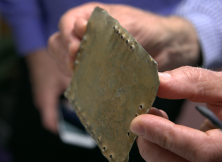
This paper reports on the materials analysis of a set of lead objects, commonly referred to as " the Jordan codices " in an attempt to shed some light on when they were manufactured. The codices are controversial, debate centring on whether they date to the early Christian period or are modern fakes. We report, for the first time, trace element analysis of samples from the lead codices as well as pieces of modern lead by PIXE and RBS to explore the trace element " fingerprint " of the metal used; the lead isotope composition by MC-ICPMS can identify the geo location of the origin of the materials used; the alpha particle emission from the lead due to the presence of the 210 Pb isotope; and the amount of helium that is trapped in the lead due to the radioactive decay of trace amounts of 238 U and 232 Th contained in the material-the (U+Th)/He age. No single technique is able to show conclusively that the objects are either modern or ancient. All four techniques suffer weaknesses, which prevent a definitive conclusion from being reached. However, there are indications that while some of the objects examined show signs of being contemporary, others appear to be older. It can be concluded that for some key examples we have been unable to show them to be made from contemporary materials and would suggest that this provides a good reason for scholars to treat the objects seriously and to perform further research on these objects.
Muonic x-ray spectroscopy uses muons to obtain information about the structure of the atom and the nucleus. In muonic atoms, the energy levels of atomic orbitals are significantly more sensitive to the finite size correction. By probing these orbitals using x-ray spectroscopy, the nuclear size correction can be extracted, providing valuable input for laser spectroscopy in the form of absolute charge radii with a relative precision better than 10−3. Continuing on developments that allowed measurements on target quantities of about 5 μg, we showed the feasibility of using implanted targets. In the future, this will allow the measurement of absolute charge radii of long-lived radioactive isotopes that are not available in sufficient enrichment or large quantities. In this contribution, we shall report on the target preparation, involving high-fluence implantation, and on the preliminary results of the muX experimental campaign.
Elemental and molecular imaging play a crucial role in understanding disease pathogenesis. To accurately correlate elemental and molecular markers, it is desirable to perform sequential elemental and molecular imaging on a single tissue section. However, very little is known about the impact of performing these measurements in sequence. In this work, we highlight some of the challenges and successes associated with performing elemental mapping in sequence with mass spectrometry imaging. Specifically, the feasibility of molecular mapping using the mass spectrometry imaging (MSI) techniques matrix assisted laser desorption ionisation (MALDI) and desorption electrospray ionisation (DESI) in sequence with the elemental mapping technique particle induced X-ray emission (PIXE) is explored. Challenges for integration include substrate compatibility, as well as delocalisation and spectral changes. We demonstrate that whilst sequential imaging comes with some compromises, sequential DESI-PIXE imaging is sufficient to correlate sulphur, iron and lipid markers in a single tissue section at the 50-micrometre scale.
The colocation of elemental species with host biomolecules such as lipids and metabolites may shed new light on the dysregulation of metabolic pathways and how these affect disease pathogeneses. Alkali metals have been the subject of extensive research, are implicated in various neurodegenerative and infectious diseases and are known to disrupt lipid metabolism. Desorption electrospray ionisation (DESI) is a widely used approach for molecular imaging, but previous work has shown that DESI delocalises ions such as potassium (K) and chlorine (Cl), precluding the subsequent elemental analysis of the same section of tissue. The solvent typically used for the DESI electrospray is a combination of methanol and water. Here we show that a novel solvent system, (50:50 (%v/v) MeOH:EtOH) does not delocalise elemental species and thus enables elemental mapping to be performed on the same tissue section post-DESI. Benchmarking the MeOH:EtOH electrospray solvent against the widely used MeOH:H2O electrospray solvent revealed that the MeOH:EtOH solvent yielded increased signal-to-noise ratios for selected lipids. The developed multimodal imaging workflow was applied to a lung tissue section containing a tuberculosis granuloma, showcasing its applicability to elementally rich samples displaying defined structural information.
Elemental imaging is widely used for imaging cells and tissues but rarely in combination with organic mass spectrometry, which can be used to profile lipids and measure drug concentrations. Here, we demonstrate how elemental imaging and a new method for spatially resolved lipidomics (DAPNe-LC-MS, based on capillary microsampling and liquid chromatography mass spectrometry) can be used in combination to probe the relationship between metals, drugs, and lipids in discrete areas of tissues. This new method for spatial lipidomics, reported here for the first time, has been applied to rabbit lung tissues containing a lesion (caseous granuloma) caused by tuberculosis infection. We demonstrate how elemental imaging with spatially resolved lipidomics can be used to probe the association between ion accumulation and lipid profiles and verify local drug distribution.
In this perspective article, we discuss the application of ion implantation to manipulate strain (by either neutralizing or inducing compressive or tensile states) in suspended thin films. Emphasizing the pressing need for a high-mobility silicon-compatible transistor or a direct bandgap group-IV semiconductor that is compatible with complementary metal-oxide-semiconductor technology, we underscore the distinctive features of different methods of ion beam-induced alteration of material morphology. The article examines the precautions needed during experimental procedures and data analysis and explores routes for potential scalable adoption by the semiconductor industry. Finally, we briefly discuss how this highly controllable strain-inducing technique can facilitate enhanced manipulation of impurity-based spin quantum bits (qubits).
An increased rate of cellular proliferation is a hallmark of cancer and may be accompanied by an increase in ribosome biogenesis and dysregulation in rRNA synthesis. In this regard, CX-5461 has been developed as a novel RNA polymerase I inhibitor and is currently in Phase I/II clinical trials for solid and hematological malignancies. In the present study, interactions between CX-5461 and single-dose X-ray exposure were assessed using isobologram analysis using MTS assay and drug-induced cell death was assessed using flow cytometric, confocal microscopy and Western blot analysis. Combination treatments involving CX-5461 and single-dose X-ray exposure highlighted increased effectiveness compared to individual treatment alone in the CaSki cervical cancer line, with marked synergistic interaction occurring within the low-drug (50 nM) and low-dose radiation range (2-6 Gy). Cell lines challenged with CX-5461 demonstrated the presence of DNA damage, induction of apoptosis, autophagy and senescence alongside high percentages of G2/M cell cycle arrest. In addition, we report preferential sensitivity of ovarian cancer cells with BRCA2 mutation to this novel agent. Taken together, CX-5461 displayed a broad spectrum of activity in a panel of solid cancer cell lines with IC50 values ranging from 35 nM to >1 mu M. The work described herein identifies the synergistic effects of CX-5461 in combination with X-rays in solid cancers and may also aid in the design of clinical trials involving this novel agent.
Ion Implantation In article number 2000237 by Nathan Cassidy, Roger Webb, David Cox, and co‐workers, preliminary results are presented from the first commercially produced implanter specifically designed for rapid and precise positioning of single ions into device structures for solid state quantum technology applications. Specifically the implantation of single bismuth ions into Si, Ge, Cu, and Au are reported, and the counting detection efficiency for single ion implants and the factors that affect such detection efficiencies are determined.
Ion irradiation and implantation have wide applications that demand accurate determination of displacement damage profile and distribution of implanted ion concentration. The prediction of vacancies is especially important to determine displacements per atom (dpa), the standard parameter of primary radiation damage in materials. However, significant discrepancies exist in estimations of vacancies between full-cascade (F-C) and quick calculation (Q-C) options in the popular computer code SRIM. This study inspected the SRIM code and a relatively new code called Iradina, which uses a similar methodology, to develop an understanding of the origin of vacancy overestimation in the F-C options for SRIM and Iradina. We found that the default values of thresholds (namely final energy in SRIM and replacement energy in Iradina) in displacement production calculations results in excessively large number of calculated vacancies and very few replacements. After conducting multiple calculations using SRIM, Iradina, and MARLOWE (all based on the binary collision approximation), a comparison of the results indicates that there is a shortcoming in the SRIM and Iradina F-C methodology for treating near-threshold collisions. This issue is responsible for the deficiency of replacements and excess of calculated vacancies in the SRIM and Iradina F-C results. Drawing on the principles of collision physics, we propose recommendations for modifying the source codes to address these issues.
LEIS depth profiles, obtained by low energy (0.5 keV) Ar+ sputtering, have been analysed for the mixed conducting oxide material La0.6Sr0.4Co0.2Fe0.8O3-δ. Samples have been examined after differing thermal treatments to examine the sub-surface reorganisation of the cation species. The profiles have shown considerable changes, but these are not strongly correlated with the thermal treatments. The similarity between the profiles suggests that preferential sputtering effects can dominate the sub-surface region (~1–3 nm) where sputtering has not reached equilibrium. Preferential sputtering of oxygen in oxide materials is well known, but here we provide evidence of the preferential sputtering of the cationic species in a complex multicomponent oxide. Of note is strong enrichment (~30%) of the sputtered surface with the heaviest of the elements, La. Simulations using the code TRIDYN have confirmed the observations, in particular, La surface enrichment and the fluence needed to achieve steady state sputtering of > 3 × 1016cm−2.
The effect of vacuum exposure on latent fingerprint chemistry has been evaluated. Fingerprints were analysed using a quartz crystal microbalance to measure changes in mass, gas chromatography mass spectrometry to measure changes in lipid composition and attenuated total reflection Fourier transform infrared spectroscopy (ATR-FTIR) to determine changes in the content of water, fatty acids and their esters after exposure to vacuum. The results are compared with samples aged under ambient conditions. It was found that fingerprints lose around 26% of their mass when exposed to vacuum conditions, equivalent to around 5 weeks ageing under ambient conditions. Further exposure to vacuum causes a significant reduction in the lipid composition of a fingerprint, in particular with the loss of tetradecanoic and pentadecanoic acid, that was not observed in ambient aged samples. There are therefore implications for sequence in which fingerprint development procedures (for example vacuum metal deposition) are carried out, as well as the use of vacuum based methods such as secondary ion mass spectrometry (SIMS) and matrix-assisted laser desorption ionisation (MALDI) in the study of fingerprint chemistry. © 2013 .
Ion Beam Analysis (IBA) consists of a set of analytical techniques addressing elemental composition of inorganic material normally conducted using ion beams in the MeV kinetic energy range. Secondary Ion Mass Spectrometry using MeV ions (MeV SIMS) is the only IBA technique which can provide extensive molecular information about organic materials. MeV ions can be extracted into air hence offering the potential to apply MeV SIMS under atmospheric pressure. At the University of Surrey Ion Beam Centre, a fully ambient MeV SIMS setup has been developed and termed “Ambient Pressure MeV SIMS”. This AP MeV SIMS can be optimized for analysis and imaging of organic molecules. MeV SIMS relies upon electronic sputtering of the target material and this is much more efficient in insulating or organic targets, and less efficient in conducting metallic materials. PIXE, on the other hand, is efficient at providing good signals from elemental metallic systems, but does not readily provide molecular information from organics. The combination of the two techniques – preferably simultaneously with the same beam – provides useful complementary information which can readily be combined. Here we present pioneering preliminary work in simultaneous molecular and elemental imaging of a complex sample comprising of two organic species and two metallic species by combining AP MeV SIMS with Heavy Ion Particle Induced X-ray emission (HIPIXE).
The proton beam writing (PBW) technique was used to fabricate microfluidic structures in SU-8 resist. A network of the buried channels was fabricated as part of a project to develop functional microfluidic device for neuronal studies and self-powered microfluidics. Protons with energies between 2.5 MeV and 0.75 MeV were used to fabricate the buried channels with a minimum feature size of around 1 μm and depths of 40–55 μm. Roughness of channels sidewalls was around 2.5 nm rms. Exposure regime and examples of functional networks fabricated using PBW are described. COMSOL Multiphysics® software was used to model the flow characteristics of fluid in the SU-8 microchannels structured by PBW. The results obtained using PBW are compared with the structures fabricated by UV-lithography.
Normal incidence argon-copper sputtering mechanisms have been investigated for ion energies just above threshold. Identical mechanisms operate in both the (111) and (001) surface orientations. Adsorption of an ordered oxygen overlay on the surface does not destroy the ejection processes. Although the mechanisms produce similar ejected atom energy distributions, the processes may be experimentally distinguishable through the angular emission spectrum of the ejected copper atoms.
We have investigated the use of conventional ion implantation to fabricate enriched 28 Si layers for use in quantum computers. The final compositions of samples enriched using ultra-low energy (800 eV and 2 keV) and low energy (20 keV) 28 Si implants of varying fluences (1x10 16-3.8x10 17 cm-2) using two different implanters were measured using channelled Rutherford Backscattering Spectroscopy. The dynamic, binary collision approximation program TRIDYN was used to model the implantation profiles to guide the analysis of the RBS spectra. It was found that ultra-low energy implants achieved high 28 Si enrichment levels but were heavily contaminated with oxygen due to poor vacuum in the implanter wafer end station. It was shown that oxidation could be reduced by using an accelerator with an end station with better vacuum and increasing the implant energy to 20 keV. However, TRIDYN simulations predict that the best 28 Si enrichment levels that could be achieved under these conditions would saturate at ~99.2 % due to self-sputtering. We modelled a range of conditions with TRIDYN and so recommend low energies (99.9 %) with the lowest possible fluences (~5-10x10 17 cm-2).
Direct analyte probed nanoextraction (DAPNe) is a technique that allows extraction of drug and endogenous compounds from a discrete location on a tissue sample using a nano capillary filled with solvent. Samples can be extracted from a spot diameters as low as 6 µm. Studies previously undertaken by our group have shown that the technique can provide good precision (5%) for analysing drug molecules in 150 µm diameter areas of homogenised tissue, provided an internal standard is sprayed on to the tissue prior to analysis. However, without an isotopically labelled standard, the repeatability is poor, even after normalisation to and the spot area or matrix compounds. By application to tissue homogenates spiked with drug compounds, we can demonstrate that it is possible to significantly improve the repeatability of the technique by incorporating a liquid chromatography separation step. Liquid chromatography is a technique for separating compounds prior to mass spectrometry (LC-MS) which enables separation of isomeric compounds that cannot be discriminated using mass spectrometry alone, as well as reducing matrix interferences. Conventionally, LC-MS is carried out on bulk or homogenised samples, which means analysis is essentially an average of the sample and does not take into account discrete areas. This work opens a new opportunity for spatially resolved liquid chromatography mass spectrometry with precision better than 20%.
This work describes the development of a new approach to measure drug levels and lipid fingerprints in single living mammalian cells. Nanocapillary sampling is an approach that enables the selection and isolation of single living cells under microscope observation. Here, live single cell nanocapillary sampling is coupled to liquid chromatography for the first time. This allows molecular species to be separated prior to ionisation and improves measurement precision of drug analytes. The efficiency of transferring analytes from the sampling capillary into a vial was optimised in this work. The analysis was carried out using standard flow liquid chromatography coupled to widely available mass spectrometry instrumentation, highlighting opportunities for widespread adoption. The method was applied to 30 living cells, revealing cell-to-cell heterogeneity in the uptake of different antibiotics. Using this system, we detected 14-158 lipid features per single cell, revealing the association between bedaquiline uptake and lipid fingerprints.
Computer simulations of ion-bombardment events which recrystallize the target demonstrate pit formation at 1.0 keV. Atoms are ejected from the first two target layers in the pit region. The remainder of the pit is created by replacement-collision sequences. Linear-cascade and spike-regime events are dynamically indistinguishable. © 1983 The American Physical Society.
This chapter describes the methods and applications for the use of multi-atom, ionized species, containing 10s to several thousand atoms, for high-flux ion doping and modification of surfaces with sub-keV energies per atom. “Massive” ion beams are formed using molecular species, liquids and gas clusters. Ion beam systems for such “massive” ions include the use of novel vaporizer methods, adiabatic cooling, electron beam ionization, and other techniques to provide stable, high-fluence beams. Applications include high-dose ion implantation for ultra-shallow junctions, room-temperature deposition of semiconductor and other layers, smoothing and high-rate etching of surfaces.
A molecular dynamics simulation has been used to investigate the sensitivity of atom ejection processes from a single-crystal target to changes in the atom-atom potential function. Four functions, three constructed from the Gibson potentials with Anderman's attractive well, and a fourth specifically developed for this investigation, were investigated in the Cu/Ar+ system over a range of ion energies from 1.0 to 10.0 kev with the KSE-B ion-atom potential. Well depths and widths also were varied. The calculations were done at normal incidence on the fcc (111) crystal orientation. Computed values were compared with experimental data where they exist. Sputtering yields, multimer yield ratios, layer yield ratios, and the ejected atom energy distribution vary systematically with the parameters of the atom-atom potential function. Calculations also were done with the modified Moliere function. Yields and other properties fall exactly into the positions predicted from the Born-Mayer function analysis. Simultaneous analysis of the ejected atom energy distribution and the ion energy dependence of the sputtering yield curve provides information about the parameters of both the wall and well portions of the atom-atom potential function.
Both Rutherford backscatterings of He-4(+) beams and non-Rutherford backscatterings of He-4(+) and H+ beams have been used in this study to investigate the depth profiles of B dopant in Mg target upon B implantation and post annealing. Primitive data analysis suggests an enhanced diffusion of surface C contaminant during the B implantation process, together with enhanced surface oxidation upon implantation and thermal annealing in flowing N-2 atmosphere. Published by Elsevier B.V.
Analysis using MeV ion beams is a thin film characterisation technique invented some 50 years ago which has recently had the benefit of a number of important advances. This review will cover damage profiling in crystals including studies of defects in semiconductors, surface studies, and depth profiling with sputtering. But it will concentrate on thin film depth profiling using Rutherford backscattering, particle induced X-ray emission and related techniques in the deliberately synergistic way that has only recently become possible. In this review of these new developments, we will show how this integrated approach, which we might call “total IBA”, has given the technique great analytical power.
The effect of 600 keV He+ ion irradiation on the temperature and magnetic field dependence of the critical current density JC in high quality BaFe1.84Co0.16As2 (Co-doped Ba122 type) thin films is investigated. The films are prepared by pulsed-laser-deposition (PLD) on CaF2 (00 $l$) substrates. The irradiation dosages are varied between 1 × 10¹³ and 1 × 1016 cm−2. Upon irradiation, the superconducting transition temperature TC drops slightly from 25 K for the unirradiated sample to about 20 K for the sample with the highest irradiation level. The JC values of the thin film samples are calculated by using the Bean critical state model. The results showed that JC could be enhanced substantially. The maximum JC value at 4.5 K temperature is enhanced up to 2.4 MA cm−2 under 1 T field. The analysis of pinning force dependence on magnetic field shows that the pinning behavior is not changed in the irradiated samples, suggesting more pinning centers of similar nature to those of presented in the unirradiated samples are introduced by the irradiation process. The results indicate that the irradiation of light element ions He+ with relatively low energy could increase the critical current density in iron based superconductors.
Molecular dynamics computer simulations have been used to study the development of ion-induced cascades in the surface region of an initially perfect single crystal metal target. A 16 mm movie has been produced to show the temporal progress of individual cascades. The cascades can then be seen to be formed from a few high energy primary knock-on initiated replacement collision sequences which overlap to form the more usual interpretation of a mature collision cascade. However, it is before the collision cascade has matured, and while the replacement sequences are spreading, that the majority of atoms (>80%) are ejected. These qualitative observations are also upheld more quantitatively in a global average, over many cascades, of the ejection time of each atom. This gives rise to the appearance of a statistical ejection front which propagates radially outwards, from the impact point on the crystal surface, with a well defined velocity. © 1984.
The suite of techniques which are available with the small accelerators used for MeV ion beam analysis (IBA) range from broad beams, microbeams or external beams using the various particle and photon spectrometries (including RBS, EBS, ERD, STIM, PIXE, PIGE, NRA and their variants), to tomography and secondary particle spectrometries like MeV-SIMS. These can potentially yield almost everything there is to know about the 3-D elemental composition of types of samples that have always been hard to analyse, given the sensitivity and the spacial resolution of the techniques used. Molecular and chemical information is available in principle with, respectively, MeV-SIMS and high resolution PIXE. However, these techniques separately give only partial information – the secret of “Total IBA” is to find synergies between techniques used simultaneously which efficiently give extra information. We here review how far “Total IBA” can be considered already a reality, and what further needs to be done to realise its full potential.
Exposure of the Hubble Space Telescope to space in low Earth orbit resulted in numerous hypervelocity impacts by cosmic dust (micrometeoroids) and anthropogenic particles (orbital debris) on the solar arrays and the radiator shield of the Wide Field and Planetary Camera 2, both subsequently returned to Earth. Solar cells preserve residues from smaller cosmic dust (and orbital debris) but give less reliable information from larger particles. Here, we present images and analyses from electron, ion and X-ray fluorescence microscopes for larger impact features (millimetre- to centimetre-scale) on the radiator shield. Validated by laboratory experiments, these allow interpretation of composition, probable origin and likely dimensions of the larger impactors. The majority (~90%) of impacts by grains greater than 50 μm in size were made by micrometeoroids, dominated by magnesium- and iron-rich silicates and iron sulfides, metallic iron-nickel and chromium-rich spinel similar to that in ordinary chondrite meteorites of asteroid origin. Our re-evaluation of the largest impact features shows substantially fewer large orbital debris impacts than reported by earlier authors. Mismatch to the NASA ORDEM and ESA MASTER models of particle populations in orbit may be partly due to model overestimation of orbital debris flux and underestimation of larger micrometeoroid numbers.
The effects of surface proximity and B concentration on end-of-range defect formation during nonmelt laser annealing in preamorphized silicon have been studied. These effects were analyzed by observing the activation and diffusion of an ultrashallow B implant, using Hall effect and secondary ion mass spectrometry measurements. By adjusting the preamorphizing implant and laser annealing conditions, B deactivation and diffusion were minimized, resulting in a sheet resistance of similar to 600 Omega/sq with a 16 nm junction depth. This is attributed to a combination of enhanced dissolution of end-of-range defects and preferential formation of B-interstitial clusters due to the surface proximity and high B concentration, respectively. (C) 2008 American Institute of Physics.
The Surrey Ion Beam Centre was awarded the Engineering and Physical Sciences Research Council (EPSRC) grant for “Promoting Cross Disciplinary Research: Engineering and Physical Sciences and Social Sciences” allowing continued research into the characteristics of desorption of secondary ions by the impact of fast primary ions in the ambient pressure at the sub-micron scale. To carry out this research a new beamline has been constructed consisting of a time-of-flight secondary ion mass spectrometer combined with the current 2MV Tandem accelerator. This research has already returned many significant results such as the first simultaneous SIMS, PIXE and RBS measurement preformed on an organic sample in vacuum. However, further optimization and validation of the new beamline is still being worked on. This work focuses on the optimization of the end station geometry to allow for high sensitivity ambient pressure measurements. It is concluded that a common geometry can be adopted for a wide variety of smooth samples to ensure optimum sensitivity provided a hard edge of the sample can be found to place the mass spectrometer capillary near.
The inherent difficulties in interpreting thermal desorption spectra are discussed. Possible mechanisms which can change the expected form of the evolution spectra are investigated including the possibility of a distribution of activation energies for release of trapped gas and the spatial distribution of the trapped gas. It is shown that the form of the evolution spectra can be quite misleading and unless care is taken the wrong conclusions can be derived. © 1979.
The introduction of strain into semiconductors offers a well-known route to modify their band structure. Here, we show a single-step procedure for generating such strains smoothly and deterministically, over a very wide range, using a simple, easily available, highly scalable, ion implantation technique to control the degree of amorphization in and around single-crystal membranes. The amorphization controls the density of the material and thus the tension in the neighboring crystalline regions. We have demonstrated up to 3.1% biaxial tensile strain and 8.5% uniaxial strain in silicon, based on micro-Raman spectroscopy. This method achieves strain levels never previously reached in mesoscopic defect-free, crystalline silicon structures. The flexible, gently controllable, single-step process points toward very high mobility complementary metal-oxide-semiconductor devices and easy fabrication of direct-bandgap germanium for silicon-compatible optoelectronics.
A molecular dynamics multiple interactions simulation computer code has been used to study the near-surface effect of cascade mixing due to ion bombardment. 90% of all moved atoms recoil forward or backward only one layer and 70% of all recoil atoms are displaced to their nearest neighbour positions. A comparison of the computed results and the results predicted from statistical theories indicates that both models would produce similar results if the lower limit in the energy threshold of the statistical theories were decreased sufficiently to include the movement of many more low energy recoils. A further comparison is made between the distributions of recoil-atom displacements at the end of the dynamic regime, and after the damaged crystallite has coalesced. There is a small degree of rearrangement, due to the collapse of collision spikes, but the resultant displacement distributions are similar. © 1983.
In this paper, the technique of microscope-spectrophotometry, used to nondestructively characterize the microstructure of ion beam synthesized iron-disilicide layers, is described. The results obtained agree extremely well, in terms of layer thickness and interfacial roughness, with those from Rutherford backscattering. The results also show that it is possible to interpret the measured spectral reflectance data in terms of: 1) defect annealing; 2) iron redistribution; and 3) phase transformations from the β to the α phase.
© 1989 Springer-Verlag Heidelberg. © 1989 Springer-Verlag Bcrbn Heidelberg. All Rights Reserved.This paper correlates photodisplacement thermal wave characterization of ion implanted silicon wafers with the lattice information provided by Rutherford Backscattering Spectrometry.
A new protocol using time-of-flight secondary ion mass spectrometry (ToF-SIMS) has been developed to identify the deposition order of a fingerprint overlapping an ink line on paper. By taking line scans of fragment ions characteristic of the ink molecules (m/z 358.2 and 372.2) where the fingerprint and ink overlap and by calculating the normalised standard deviation of the intensity variation across the line scan, it is possible to determine whether or not a fingerprint is above ink on a paper substrate. The protocol adopted works for a selection of fingerprints from four donors tested here and for a fingerprint that was aged for six months; for one donor, the very faint fingerprints could not be visualized using either standard procedures (ninhydrin development) or SIMS and therefore the protocol correctly gives an inconclusive result.
The theoretical error rates in deterministic ion implantation when using an ion beam governed by a Poisson point process with a detector that counts the impacts are investigated. It is concluded that if the error rates are small, then for spots with nominally one implanted ion the probability of failure to implant the correct number is ≈ 𝜅/𝜆+𝜂⎯⎯+𝜆/2 for a synchronous (i.e., pulsed) system or 𝐾/𝐿+𝜂⎯⎯+𝐿𝑡s for an asynchronous (i.e., continuous beam) system, where 𝜂⎯⎯ is the probability that the detector misses an ion impact, and 𝐿(𝐾) and 𝜆(𝜅) are the number of ions (dark counts) per unit time and per pulse, respectively. ts is the system reaction time for an asynchronous system. This approximation allows easy identification of the greatest need for engineering effort. Some experimental efforts to measure these parameters and their uncertainties are examined.
BACKGROUND: Recent publications have explored the possibility of using fingerprints to confirm drug use, but none has yet dealt with environmental contamination from fingertips. Here we explored the possibility of establishing an environmental cutoff for drug testing from a single fingerprint. METHODS: Fingerprint samples (n=100) were collected from the hands of 50 nondrug users before and after handwashing to establish separate environmental cutoff values and testing protocols for cocaine, benzoylecgonine, heroin, and 6-monoacetylmorphine. The cutoff was challenged by testing the fingerprints of drug-free volunteers after shaking hands with drug users. Fingerprints from patients who testified to taking cocaine (n = 32) and heroin (n = 24) were also collected and analyzed. RESULTS: A different cutoff value needed to be applied, depending on whether the fingerprints were collected as presented or after handwashing. Applying these cutoffs gave a 0%false-positive rate from the drug-free volunteers. After application of the cutoff, the detection rate (compared to patient testimony) for washed hands of patients was 87.5% for cocaine use and 100% for heroin use. CONCLUSIONS: Fingerprints show enhanced levels of cocaine, heroin, and their respective metabolites in patients who testified to taking the substances, compared with the population of naı¨ve drug users surveyed, and a cutoff (decision level) can be established. The cutoff is robust enough to account for small increases in analyte observed after secondary transfer.
This book is an introduction to the application of computer simulation and theory in the study of the interaction of energetic particles (1 ev to the MeV range) with solid surfaces. The authors describe methods that are applicable both to hard collisions between nuclear cores of atoms down to soft interactions, where chemical effects or long-range forces dominate. The range of potential applications of the technique is enormous. In surface science, applications include surface atomic structure determination using ion scattering spectroscopy or element analysis using SIMS or other techniques that involve depth profiling. Industrial applications include optical or hard coating deposition, ion implantation in semiconductor device manufacture or nanotechnology. The techniques described will facilitate studying plasma-sidewall interaction in fusion devices. This book will be of interest to graduate students and researchers, both academic and industrial, in surface science, semiconductor engineering, thin-film deposition and particleSHsurface interactions in departments of physics, chemistry and electrical engineering.
Ultra-shallow B and BF 2 implants in silicon pre-amorphised with Ge have been activated using a scanning non-melt laser. The implants were activated either by using 1 or 10 laser scans. Isochronal 60s post-laser annealing between 700-1000°C were then undertaken to study the deactivation and reactivation of the B. Both B and BF2 samples were implanted with a dose of 1×10 15 B cm -2 at an effective energy of 500eV. The presence of F from the BF 2 implants, which is superimposed over the boron profile increases the sheet resistance of the initial fabricated junction (from 600-700 ohms/sq from B implants only to 750-1100 ohms/sq for BF2 implants). Fluorine also changes the deactivation and reactivation behaviour of the boron during the post-anneals by increasing the amount of deactivation of the boron. © 2006 Materials Research Society.
A new geometrical theory of sputtering is extended to investigate the behaviour of interface mixing as a function of the angle of ion incidence. It is found that the amount of mixing peaks with the angle of incidence at different angles depending upon the depth of the interface. When the interface is at the surface the mixing parameter behaves as the sputtering yield and maximises around 70 degree , whilst deeper interfaces, around the damage range, have maximum mixing at normal incidence.
Characterizing proton beam damage in biological materials is of interest to enable the integration of proton microprobe elemental mapping techniques with other imaging modalities. It is also of relevance to obtain a deeper understanding of mechanical damage to lipids in tissues during proton beam cancer therapy. We have developed a novel strategy to characterize proton beam damage to lipids in biological tissues based on mass spectrometry imaging. This methodology is applied to characterize changes to lipids in tissues ex vivo, irradiated under different conditions designed to mitigate beam damage. This work shows that performing proton beam irradiation at ambient pressure, as well as including the application of an organic matrix prior to irradiation, can reduce damage to lipids in tissues. We also discovered that, irrespective of proton beam irradiation, placing a sample in a vacuum prior to desorption electrospray ionization imaging can enhance lipid signals, a conclusion that may be of future benefit to the mass spectrometry imaging community.
The use of polyatomic (cluster) ion beams for SIMS has proven to be an efficient method for the characterization of solids. Computer simulation programs such as Molecular Dynamics (MD) are often run to gain an insight into the ion-solid interactions that take place under these circumstances; however, for simulations to be able to make accurate predictions, a massive amount of computational resources are required to be at hand. These include several months in simulation time and the use of very large targets, not to mention that a single simulation run is a representation of only a single ion trajectory. Thus, MD simulations are invaluable for gaining insight into ion-solid interactions but are less so for being able to provide information when time constraints are put in place. The work here aims to achieve a prediction model that, when completely functional, will be able to deal with the pressures of the clock. This, we believe, is achievable by employing simpler modeling criteria that are dependant upon aspects of an initial energy deposition profile within the target under irradiation. Copyright © 2010 John Wiley & Sons, Ltd.
Multiple interaction computer simulations of bombardment cascades have been used to study the surface damage created by an ion. Qualitative information on the temporal evolution of the dynamic cascade is obtained from an animated movie of the process. A comparison is made between the final damage states of high energy-density trajectories. A number of ion-atom potential functions have been investigated over a wide range of ion energies. Only superficial differences are found. © 1984.
Secondary Ion Mass spectrometry (SIMS) has been generally used in the field of material sciences. In recent years, it has also been applied for molecular imaging of biological samples. Nevertheless, molecular ions derived from the large molecules (more than 1 kDa) were detected with very low sensitivity. Plasma desorption mass spectrometry (PDMS) is known as mass spectrometry for large organic molecule. In PDMS, fission fragments bombard samples and the impact induces molecular ionization by electronic excitation. Large organic molecules are detected by using swift heavy ions in SIMS. In this work, 6 MeV Cu4+ we irradiated angiotensin II, a class of peptides. The intact molecular ions generated by swift heavy ion irradiation were analyzed by time‐of‐flight (TOF) measurement. The yields are compared with some other probe ions, bismuth or flurane. Swift heavy ion bombardment ionized large organic molecules more effectively than other probes. Therefore, high energy ion can be applied in high resolution molecular imaging.
The International Roadmap for Semiconductors requires ultrashallow, highly activated, abrupt dopant profiles in the source/drain extension regions, for technology nodes beyond 45 nm. The authors contrast B and BF2 implants in Si and silicon on insulator (SOI) substrates with and without a preamorphizing implant (PAI). The objective of the study is to compare between Si and SOI substrates, PAI and non-PAI condition, and B and BF2 implants. The results show the absence of the "reverse annealing effect" in BF2 implants, which is observed in B implants. The presence of F appears to impede the formation of boron interstitial clusters, which is shown in the case of B implant. The BF2 implants follow a similar trend for SOI and Si with and without PAI. (C) 2008 American Vacuum Society.
The spatial variation of energy deposited in a solid can lead to local variations in sputtering yield at points on the surface neighbouring the point of ion impact. An approximate theory is developed to describe this local sputtering yield variation in terms of the local morphology. It is then shown how, if this local variation merely moderates the standard sputtering yield-projectile incidence angle function by multiplication, an erosion slowness theory can be simply modified and generalized to allow prediction of the time development of sputtered surface morphology. Both transient and steady-state morphologies are explored. © 1981 Chapman and Hall Ltd.
The creation of stable, highly conductive ultrashallow junctions in strained Si is a key requirement for future Si based devices. It is shown that in the presence of tensile strain, Sb becomes a strong contender to replace As as the dopant of choice due to advantages in junction depth, junction steepness, and crucially, sheet resistance. While 0.7% strain reduces resistance for both As and Sb, a result of enhanced electron mobility, the reduction is significantly larger for Sb due to an increase in donor activation. Differential Hall and secondary-ion mass spectroscopy measurements suggest this to be a consequence of a strain-induced Sb solubility enhancement following epitaxial regrowth, increasing Sb solubility in Si to levels approaching 10(21) cm(-3). Advantages in junction depth, junction steepness, and dopant activation make Sb an interesting alternative to As for ultrashallow doping in strain-engineered complementary metal-oxide semiconductor devices. (c) 2008 American Vacuum Society.
Ambient Pressure MeV SIMS (AP MeV SIMS) is a special application of MeV SIMS technique for molecular detection and imaging under ambient conditions. There are several advantages of using ambient over nonambient techniques such as minimising or completely avoiding sample preparation that can contribute to the reducing of costs and shortening of analysis. Moreover, by performing analysis in ambient conditions negative vacuum influence on samples will be avoided. The emergence of ambient mass spectrometry techniques over the past decade has been enormous with a broad range of applications such as food quality, environmental analysis and life sciences [1,2]. On the other hand, the disadvantage of ambient pressure mass spectrometry techniques is the influence of the ambient background which can suppress the signal from the target. In AP MeV SIMS, molecular species present in ambient surrounding of the sampling site will also be ionised by ion beams hence secondary ions originating from the sample have to be transported in the most efficient manner from the site of interaction of ion beams and target into the mass spectrometer capillary and, finally, into the mass spectrometer [3]. In this work, we present the optimisation of the mass spectrometer capillary temperature, distances of mass spectrometer and sheath gas, in our case helium, with respect to the beam axis and angle of the helium flow capillary with respect to the sample.
The MIAMI* facility at the University of Huddersfield is one of a number of facilities worldwide that permit the ion irradiation of thin foils in-situ in a transmission electron microscope. MIAMI has been developed with a particular focus on enabling the in-situ implantation of helium and hydrogen into thin electron transparent foils, necessitating ion energies in the range 1 - 10 keV. In addition, however, ions of a variety of species can be provided at energies of up to 100 keV (for singly charged ions), enabling studies to focus on the build up of radiation damage in the absence or presence of implanted gas. This paper reports on a number of ongoing studies being carried out at MIAMI, and also at JANNuS (Orsay, France) and the IVEM / Ion Accelerator Facility (Argonne National Lab, US). This includes recent work on He bubbles in SiC and Cu; the former work concerned with modification to bubble populations by ion and electron beams and the latter project concerned with the formation of bubble super-lattices in metals. A study is also presented consisting of experiments aimed at shedding light on the origins of the dimensional changes known to occur in nuclear graphite under irradiation with either neutrons or ions. Single crystal graphite foils have been irradiated with 60 keV Xe ions in order to create a non-uniform damage profile throughout the foil thickness. This gives rise to varying basal-plane contraction throughout the foil resulting in almost macroscopic (micron scale) deformation of the graphite. These observations are presented and discussed with a view to reconciling them with current understanding of point defect behavior in graphite.*Microscope and Ion Accelerator for Materials Investigations Copyright © Materials Research Society 2014.
Fingerprints have been proposed as a promising new matrix for drug testing. In previous work it has been shown that a fingerprint can be used to distinguish between drug users and non-users. Herein, we look at the possibility of using a fingerprint to distinguish between dermal contact and administration of heroin. Fingerprint samples were collected from (a) 10 patients attending a drug rehabilitation clinic (b) 50 non-drug users (c) participants who touched 2 mg street heroin, before and after various hand cleaning procedures. Oral fluid was also taken from the patients. All samples were analysed using a liquid chromatography – high resolution mass spectrometry (LC-HRMS) method validated in previous work for heroin and 6-AM. The HRMS data was analysed retrospectively for morphine, codeine, 6-acetylcodeine and noscapine. Heroin and 6-AM were detected in all fingerprint samples produced from contact with heroin, even after handwashing. In contrast, morphine, acetylcodeine and noscapine were successfully removed after handwashing. In patient samples, the detection of morphine, noscapine and acetylcodeine (alongside heroin and 6-AM) gave a closer agreement to patient testimony on whether they had recently used heroin use than the detection of heroin and 6-AM alone. This research highlights the importance of washing hands prior to donating a fingerprint sample to distinguish recent contact with heroin from heroin use.
A molecular dynamics simulation has been used to study the times at which atoms are ejected during sputtering events. Plots of the atom ejection time versus distance from the impact point indicate that many sputtering events occur along a roughly circular front that propagates outward at hypersonic speed.
Surface mass spectrometry methods can be difficult to use effectively with low cost, portable mass spectrometers. This is because commercially available portable (single quadrupole) mass spectrometers lack the mass resolution to confidently differentiate between analyte and background signals. Additionally, current surface analysis methods provide no facility for chromatographic separation and therefore are vulnerable to ion suppression. Here we present a new analytical method where analytes are extracted from a sample using a solvent flushed across the surface under high pressure, separated using a chromatography column and then analysed using a portable mass spectrometer. The use of chromatography reduces ion suppression effects and this, used in combination with in-source fragmentation, increases selectivity, thereby allowing high sensitivity to be achieved with a portable and affordable quadrupole mass spectrometer. We demonstrate the efficacy of the method for the quantitative detection of cocaine and benzoylecgonine in urine and oral fluid. The method gives relative standard deviations below 15% (with one exception), and R2 values above 0.998. The limits of detection for these analytes in oral fluid and urine are
Computer simulation of ion-bombardment events has been extended to coalesce and recrystallise the target, so that the surface damage created when a single ion strikes a metal surface can now be studied directly. Qualitative and quantitative information on pit formation, other surface damage and recoil mixing can be obtained from the model. Many characteristics of the target's final condition are evident before the excess energy has been removed. Small structured and faceted surface pits are found at 1.0 keV ion energy for both low and high energy-density potential functions. Atoms are only sputtered from the first two layers of the pit region. The rest of the pit is formed by atoms displaced laterally or downward, initiating replacement collision sequences. A movie has been produced which presents the temporal development and creation of surface damage, and demonstrates surface reconstruction processes. © 1983.
Return of materials from the Hubble Space Telescope (HST) during shuttle orbiter service missions has allowed inspection of large numbers of hypervelocity impact features from long exposure at about 615 km altitude in low Earth orbit (LEO) [1,2]. Here we describe the application of advanced X-ray microanalysis techniques on scanning electron microscopes (SEM), microprobes and a 2 MV Tandetron, to nearly 400 impacts on the painted metal surface of the Wide Field and Planetary Camera 2 (WFPC2) radiator shield [3,4]. We identified artificial Orbital Debris (OD) and natural Micrometeoroid (MM) origins for small [5] and even for larger particles [6], which usually may leave little or no detectable trace on HST solar arrays, as they penetrate through the full cell thickness [2,7].
An apparatus has been built to perform irradiation and electrical testing of REBCO coated conductors (CC) held below their critical temperature (Tc). Patterned tracks of Fujikura GdBCO CC were irradiated with 2 MeV He+ ions in steps up to 4 mdpa whilst held at 40 K, and the critical current density (Jc) determined from I–V characteristics. These 'in-situ' samples then underwent annealing experiments at room temperature. The superconducting performance, both before and after room temperature annealing, has been compared to equivalent samples irradiated at room temperature and then cooled for testing at 40 K to understand how the damage tolerance of these materials is affected by sample temperature. Details of the apparatus and experimental results from preliminary work are presented and discussed. These preliminary results show that both Tc and Jc values of patterned tracks degrade with irradiation dose, with most samples showing similar behaviour. The room temperature annealing of 'in-situ' irradiated samples resulted in a significant recovery of properties. We conclude that irradiation temperature does alter how the superconducting properties of GdBCO CC are affected by ion irradiation, and that this observation has implications for the design of high temperature superconducting magnets for future fusion reactors.
Transmission electron microscopy (TEM) of ex-situ He ion irradiated bulk W has been performed to quantitatively compare the damage microstructure to that observed in regions of comparable thicknesses during in-situ ion irradiation with TEM experiments. Samples were irradiated to achieve He-appm/DPA ratios of 2000 and 500 at temperatures of 500 and 800 °C to 1.5 and 3.0 DPA. For irradiations at 500 °C, bubble diameters (∼2 nm) were larger and areal bubble densities (∼1012 bubbles/cm2) were lower than those in the in-situ experiments. This is attributed to greater amounts of He being retained in the ex-situ bulk experiments whereas in the in-situ experiments some may escape due to the proximity of surfaces. Dislocation loops were observed in all samples and were characterised as b = ±½ type with no b = type loops. Dislocation loop populations were dominated by interstitial type (∼60%) agreeing with in-situ experiments. However, dislocation loops in this work were larger, ranging in size from 7 to 100 nm and large concentrations of entangled dislocation lines were observed in the bulk of the grain as compared to in the in-situ experiments.
Direct analyte-probed nano-extraction (DAPNe) is a method of extracting material from a microscale region of a sample and provides the opportunity for detailed mass spectrometry analysis of extracted analytes from a small area. The technique has been shown to provide enhanced sensitivity compared with bulk analysis by selectively removing analytes from their matrix and has been applied for selective analysis of single cells and even single organelles. However, the quantitative capabilities of the technique are yet to be fully evaluated. In this study, various normalisation techniques were investigated in order to improve the quantitative capabilities of the technique. Two methods of internal standard incorporation were applied to test substrates, which were designed to replicate biological sample matrices. Additionally, normalisation to the extraction spot area and matrix compounds were investigated for suitability in situations when an internal standard is not available. The variability observed can be significantly reduced by using a sprayed internal standard, and in some cases, by normalising to the extracted area.
BACKGROUND: Paper spray mass spectrometry6 is a technique that has recently emerged and has shown excellent analytical sensitivity to a number of drugs in blood. As an alternative to blood, fingerprints have been shown to provide a noninvasive and traceable sampling matrix. Our goal was to validate the use of fingerprint samples to detect cocaine use. METHODS: Samples were collected on triangular pieces (168 mm2) of washed Whatman Grade I chromatography paper. Following application of internal standard, spray solvent and a voltage were applied to the paper before mass spectrometry detection. A fingerprint visualization step was incorporated into the analysis procedure by addition of silver nitrate solution and exposing the sample to ultraviolet light. RESULTS: Limits of detection for cocaine, benzoylecgonine, and methylecgonine were 1, 2, and 31 ng/mL respectively, with relative standard deviations of less than 33%. No matrix effects were observed. Analysis of 239 fingerprint samples yielded a 99% true-positive rate and a 2.5% false-positive rate, based on the detection of cocaine, benzoylecgonine, or methylecgonine with use of a single fingerprint. CONCLUSIONS: The method offers a qualitative and noninvasive screening test for cocaine use. The analysis method developed is rapid (4 min/sample) and requires no sample preparation.


