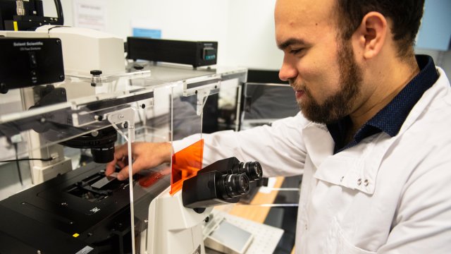
Dr Rita Jabr


Senior Lecturer in Cardiac Electrophysiology
+44 (0)1483 684618
12 AY 04

About

University roles and responsibilities

  • Senior Lecturer in Cardiac Electrophysiology

    Previous roles

    Lecturer in Cardiac Electrophysiology
    University of Surrey
    Senior Research Fellow
    Department of Cardiac Electrophysiology, National Heart and Lung Institute, Imperial College London
    Senior Research Fellow
    Cardiovascular Division, Department of Cardiology, The Rayne Institute, King's College London
    Honorary Lecturer/Senior Research Fellow
    Centre for Clinical Pharmacology and Toxicology, Department of Medicine, University College London
    Assistant Professor
    Department of Physiology, Faculty of Medicine, Kuwait University
    Postdoctoral Fellow
    Department of Physiology and Cell Biology, School of Medicine, University of Nevada
    PhD Student
    Physiology Department, University of Manitoba

    Affiliations and memberships

    Member
    AES (Qatar Foundation's premier intellectual exchange network and Arab science revival initiative)
    Member
    British Society for Cardiovascular Research
    Member
    International Society for Heart Research
    Member
    Physiological Society UK
    Member
    Royal Society of Medicine

    Research

    Research interests

    Research collaborations

    Technical skills

    Electrophysiology

    • Patch-clamp technique
    • Intracellular microelectrode or extracellular monophasic action potential recording simultaneously with isometric muscle contractions
    • Tissue impedance measurement
    • Multi-electrode array system
    • Dielectrophoresis

    Muscle contraction

    • Recording isometric muscle contractions in cardiac and smooth muscle preparations
    • Measurement of isolated cardiomyocyte mechanics

    Imaging techniques

    • Immunofluorescence with laser scanning confocal microscopy
    • Real-time intracellular Ca2+ imaging by epifluorescence microscopy, cell shortening by video-scanning methods

    Biochemistry

    • Protein phosphatase activity assays
    • Subcellular tissue fractionation
    • SDS-PAGE and western blotting
    • PCR
    • Mouse genotyping
    • Baculovirus protein expression system.

    Cell preparation

    • Single cardiac and vascular smooth muscle cells isolation techniques

    Tissue culture

    • Primary and cell-line tissue culture
    • Proliferation assay

    Funding

    • 2013-2015 (£128,000; PI: R Jabr, Co-I: CH Fry). Title: Investigation of the role of calcineurin in persistent electrophysiological myocardial changes following regression from left ventricular hypertrophy. Funded by British Heart Foundation
    • 2013-2014 (£8,000; Co-PI: R Jabr). Title: MicroRNAs as potential biomarkers in coronary artery calcification. Biomedical & Health Research Pump-Priming Funds grant, University of Surrey, 2011-2015 (£638,000) HASTE Foundation. Title: Improving detection and characterising the fundamental basis of atrial fibrillation. I am the Lead on the investigative research component (Dr E Leatham is lead on the clinical component and Prof CH Fry the manager of both components)
    • 2008-2010 (£147,280; PI: R Jabr, Co-I: CH Fry). Title: The role of calcineurin in regulating action potential propagation in normal and hypertrophied myocardium through connexin43 dephosphorylation. Funded by British Heart Foundation
    • 2009-2012 (€300,000; PI: CH Fry; Co-I, R Jabr). Title: Combatting Incontinence: InComb-EU Framework 7. 
    • 1999-2000 (£80,000- PI R Jabr). Title: Contractile and electrophysiological properties of cardiac ventricular myocytes in spontaneously hypertensive rats before and during development of hypertension, 1999. Funded by Kuwait University.

    Invited research lectures

    • 2012 Centre for Cardiovascular investigation; Faculty of Medicine; University of La Plata. Title: The differential role of calcineurin isoforms in the cardiovascular system.
    • 2011 Physiological Society Meeting, Oxford, July, 2011 Title: Inter-relationship between conduction velocity, intracellular Ca2+ and gap junction resistance in ventricular myocardium
    • 2011 University of Surrey, FHMS, research day, 'Hot topics in cardiovascular research'. Title: Cellular mechanisms for cardiac arrhythmogenesis.
    • 2010 Pfizer, sandwich, UK. Title: Role of calcineurin in overactive bladder.
    • 2010 The FP7 mid-term meeting, UK. Title: Use of the dielectrophoresis technique for selective cell sorting of bladder urothelial cells.
    • 2009 The European Society of Cardiology, Munich, Germany. Title: Calcineurin nuclear import.
    • 2009 The Marie Curie- Nuclear Receptors Biology Meeting, UK. Title: Role of nuclear calcineurin in the heart.
    • 2008 UK Gap Junction meeting, University of Surrey, UK. Title: Calcineurin slows conduction velocity during rapid pacing in guinea pig papillary muscle.

    Supervision

    Postgraduate research supervision

    Completed postgraduate research projects I have supervised

    Teaching

    Publications

    Navroop Johal, Kevin Cao, Callum Arthurs, Michael Millar, Christopher Thrasivoulou, Aamir Ahmed, Rita I Jabr, Dan Wood, Peter Cuckow, Christopher H Fry (2021)Contractile function of detrusor smooth muscle from children with posterior urethral valves – The role of fibrosis, In: Journal of Pediatric Urology17(1)pp. 100.e1-100.e10 Elsevier Ltd

    Introduction Posterior urethral valves (PUV) is the most common cause of congenital bladder outflow obstruction with persistent lower urinary tract and renal morbidities. There is a spectrum of functional bladder disorders ranging from hypertonia to bladder underactivity, but the aetiology of these clinical conditions remains unclear. Aims and objectives We tested the hypothesis that replacement of detrusor muscle with non-muscle cells and excessive deposition of connective tissue is an important factor in bladder dysfunction with PUV. We used isolated detrusor samples from children with PUV and undergoing primary or secondary procedures in comparison to age-matched data from children with functionally normal bladders. In vitro contractile properties, as well as passive stiffness, were measured and matched to histological assessment of muscle and connective tissue. We examined if a major pathway for fibrosis was altered in PUV tissue samples. Methods Isometric contractions were measured in vitro in response to either stimulation of motor nerves to detrusor or exposure to cholinergic and purinergic receptor agonists. Passive mechanical stiffness was measured by rapid stretching of the tissue and recording changes to muscle tension. Histology measured the relative amounts of detrusor muscle and connective tissue. Multiplex quantitative immunofluorescence labelling using five epitope markers was designed to determine cellular pathways, in particular the Wnt-signalling pathway, responsible for any changes to excessive deposition of connective tissue. Results and Discussion PUV tissue showed equally reduced contractile function to efferent nerve stimulation or exposure to contractile agonists. Passive muscle stiffness was increased in PUV tissue samples. The smooth muscle:connective tissue ratio was also diminished and mirrored the reduction of contractile function and the increase of passive stiffness. Immunofluorescence labelling showed in PUV samples increased expression of the matrix metalloproteinase, MMP-7; as well as cyclin-D1 expression suggesting cellular remodelling. However, elements of a fibrosis pathway associated with Wnt-signalling were either reduced (β-catenin) or unchanged (c-Myc). The accumulation of extracellular matrix, containing collagen, will contribute to the reduced contractile performance of the bladder wall. It will also increase tissue stiffness that in vivo would lead to reduced filling compliance. Conclusions Replacement of smooth muscle with fibrosis is a major contributory factor in contractile dysfunction in the hypertonic PUV bladder. This suggests that a potential strategy to restore normal contractile and filling properties is development of the effective use of antifibrotic agents.

    Vadim Alexeenko, Philippa J Howlett, James A Fraser, Daniel Abasolo, Thang S Han, David S Fluck, Christopher H Fry, Rita I Jabr (2021)Prediction of Paroxysmal Atrial Fibrillation From Complexity Analysis of the Sinus Rhythm ECG: A Retrospective Case/Control Pilot Study, In: Frontiers in physiology12pp. 570705-570705 Frontiers Media S.A

    Paroxysmal atrial fibrillation (PAF) is the most common cardiac arrhythmia, conveying a stroke risk comparable to persistent AF. It poses a significant diagnostic challenge given its intermittency and potential brevity, and absence of symptoms in most patients. This pilot study introduces a novel biomarker for early PAF detection, based upon analysis of sinus rhythm ECG waveform complexity. Sinus rhythm ECG recordings were made from 52 patients with ( n = 28) or without ( n = 24) a subsequent diagnosis of PAF. Subjects used a handheld ECG monitor to record 28-second periods, twice-daily for at least 3 weeks. Two independent ECG complexity indices were calculated using a Lempel-Ziv algorithm: R-wave interval variability (beat detection, BD) and complexity of the entire ECG waveform (threshold crossing, TC). TC, but not BD, complexity scores were significantly greater in PAF patients, but TC complexity alone did not identify satisfactorily individual PAF cases. However, a composite complexity score ( h -score) based on within-patient BD and TC variability scores was devised. The h -score allowed correct identification of PAF patients with 85% sensitivity and 83% specificity. This powerful but simple approach to identify PAF sufferers from analysis of brief periods of sinus-rhythm ECGs using hand-held monitors should enable easy and low-cost screening for PAF with the potential to reduce stroke occurrence.

    Kai Hoettges, Erin Henslee, Ruth M. Torcal Serrano, Rita Jabr, Rula Abdallat, Andrew Beale, Abdul Waheed, Patrizia Camelliti, Christopher Fry, Daan R. van der Veen, Fatima Labeed, Michael Hughes (2019)Ten–Second Electrophysiology: Evaluation of the 3DEP Platform for high-speed, high-accuracy cell analysis., In: Scientific Reports919153 Nature Research

    Electrical correlates of the physiological state of a cell, such as membrane conductance and capacitance, as well as cytoplasm conductivity, contain vital information about cellular function, ion transport across the membrane, and propagation of electrical signals. They are, however, difficult to measure; gold-standard techniques are typically unable to measure more than a few cells per day, making widespread adoption difficult and limiting statistical reproducibility. We have developed a dielectrophoretic platform using a disposable 3D electrode geometry that accurately (r2 > 0.99) measures mean electrical properties of populations of ~20,000 cells, by taking parallel ensemble measurements of cells at 20 frequencies up to 45 MHz, in (typically) ten seconds. This allows acquisition of ultra-high-resolution (100-point) DEP spectra in under two minutes. Data acquired from a wide range of cells – from platelets to large cardiac cells - benchmark well with patch-clamp-data. These advantages are collectively demonstrated in a longitudinal (same-animal) study of rapidly-changing phenomena such as ultradian (2–3 hour) rhythmicity in whole blood samples of the common vole (Microtus arvalis), taken from 10 µl tail-nick blood samples and avoiding sacrifice of the animal that is typically required in these studies.

    M Pakzad, Y Ikeda, C McCarthy, DG Kitney, Rita Jabr, Christopher Fry (2016)Contractile effects and receptor analysis of adenosine-receptors in human detrusor muscle from stable and neuropathic bladders, In: Naunyn-Schmiedeberg's Archives of Pharmacology389(8)pp. 921-929 Springer

    To measure the relative transcription of adenosine receptor subtypes and the contractile effects of adenosine and selective receptor-subtype ligands on detrusor smooth muscle from patients with neuropathic overactive (NDO) and stable bladders and also from guinea-pigs. Contractile function was measured at 37°C in vitro from detrusor smooth muscle strips. Contractions were elicited by superfusate agonists or by electrical field stimulation. Adenosine-receptor (A1, A2A, A2B, A3) transcription was measured by RT-PCR. Adenosine attenuated nerve-mediated responses with equivalent efficacy in human and guinea-pig tissue (pIC50 3.65–3.86); the action was more effective at low (1–8 Hz) compared to high (20– 40 Hz) stimulation frequencies in human NDO and guinea-pig tissue. With guinea-pig detrusor the action of adenosine was mirrored by the A1/A2-agonist N-ethylcarboxamido adenosine (NECA), partly abolished in turn by the A2B-selectve antagonist alloxazine, as well as the A1-selective agonist N6- cyclopentyladenosine (CPA). With detrusor from stable human bladders the effects of NECA and CPA were much smaller than that of adenosine. Adenosine also attenuated carbachol contractures, but mirrored by NECA (in turn blocked by alloxazine) only in guinea-pig tissue. Adenosine receptor subtype transcription was measured in human detrusor and was similar in both groups, except reduced A2A levels in overactive bladder. Suppression of the carbachol contracture in human detrusor is independent of A-receptor activation, in contrast to an A2B-dependent action with guinea-pig tissue. Adenosine also reduced nerve-mediated contractions, by an A1- dependent action suppressing ATP neurotransmitter action.

    Bahareh Vahabi, RITA JABR, Chris Fry, Karen McCloskey, Karel Everaert, Christina W. Agudelo, Thomas F. Monaghan, Mohammad S. Rahnama'i, Jalesh N. Panicker, Jeffrey P. Weiss (2020)ICI‐RS 2019 nocturia think tank: How can experimental science guide us in understanding the pathophysiology of nocturia?, In: Neurourology and Urodynamics39(S3) Wiley

    Introduction The following is a report on the proceedings of the 2019 International Consultation on Incontinence‐Research Society nocturia think tank (NTT). Objectives The objectives of the 2019 NTT were as follows: (a) to evaluate the role of urothelium in the pathophysiology of nocturia; (b) to determine whether nocturia is a circadian disorder; (c) to discuss the role of melatonin in nocturia; (d) to consider ambulatory urodynamic monitoring in evaluating patients with nocturia; (e) to explore studies of water handling in human compartments utilizing heavy water; and (f) to explore whether basic science is the key to understanding the treatment options for diminished bladder capacity in patients with nocturia. Methods A compendium of discussions of the role of experimental science in understanding the pathophysiology of nocturia is described herein. Results and Conclusions Translational science will play an increasing role in understanding the pathophysiology of nocturia, which may result in improved treatment strategies.

    Christopher Henry Fry, Basu Chakrabarty, Hikaru Hashitani, Karl‐Erik Andersson, Karen McCloskey, RITA JABR, Marcus J. Drake (2020)New targets for overactive bladder—ICI‐RS 2109, In: Neurourology and Urodynamics39(S3) Wiley

    Aim To review evidence for novel drug targets that can manage overactive bladder (OAB) symptoms. Methods A think tank considered evidence from the literature and their own research experience to propose new drug targets in the urinary bladder to characterize their use to treat OAB. Results Five classes of agents or cellular pathways were considered. (a) Cyclic nucleotide–dependent (cyclic adenosine monophosphate and cyclic guanosine monophosphate) pathways that modulate adenosine triphosphate release from motor nerves and urothelium. (b) Novel targets for β3 agonists, including the bladder wall vasculature and muscularis mucosa. (c) Several TRP channels (TRPV1, TRPV4, TRPA1, and TRPM4) and their modulators in affecting detrusor overactivity. (d) Small conductance Ca2+‐activated K+ channels and their influence on spontaneous contractions. (e) Antifibrosis agents that act to modulate directly or indirectly the TGF‐β pathway—the canonical fibrosis pathway. Conclusions The specificity of action remains a consideration if particular classes of agents can be considered for future development as receptors or pathways that mediate actions of the above mentioned potential agents are distributed among most organ systems. The tasks are to determine more detail of the pathological changes that occur in the OAB and how the specificity of potential drugs may be directed to bladder pathological changes. An important conclusion was that the storage, not the voiding, phase in the micturition cycle should be investigated and potential targets lie in the whole range of tissue in the bladder wall and not just detrusor.

    CH Fry, RI Jabr (2010)The action potential and nervous conduction, In: Surgery28(2)pp. 49-54 Elsevier

    An actionpotential is a transient depolarization of the membrane potential of excitable cells. They serve two main functions: to transmit and encode information, and to initiate cellular events such as muscular contraction. In this article actionpotentials generated in nerves will be the focus of attention. An actionpotential results from a transient change to the properties of the cell membrane, from a state where it is much more permeable to K+ than Na+, to a reversal of these permeability properties. Thus during the actionpotential an influx of Na+ is responsible for the rapid depolarization and an efflux of K+ causes repolarization. This ionic basis of the actionpotential can be predicted from the Nernst equation and is illustrated in the text. Changes to membrane ionic permeability are due to the opening and closing of voltage-gated ion channels, and the properties of such channels explain additional phenomena such as refractoriness, threshold and cellular excitability. Actionpotentials conduct with a finite velocity along nerve axons, and the actual velocity depends on a number of factors that include: fibre radius, temperature, functional ion channel number and the presence of a myelin sheath. The physical basis of conduction is explained by the local circuit hypothesis. Synaptic transmission of an actionpotential is explained in terms of excitatory post-synaptic potential (EPSP) generation at the post-synaptic membrane. The facility by which post-synaptic actionpotential may be developed is explained in terms of temporal and spatial summation as well as the influence of inhibitory transmitters.

    Carly J. McCarthy, Youko Ikeda, Deborah Skennerton, Basu Chakrabarty, Anthony J. Kanai, Rita I. Jabr, Christopher H. Fry (2019)Characterisation of nerve‐mediated ATP release from detrusor muscle; pathological implications, In: British Journal of Pharmacology Wiley

    Background and Purpose. To characterise the molecular mechanisms that determine variability of atropine‐resistance of nerve‐mediated contractions in human and guinea‐pig detrusor smooth muscle Experimental Approach. Atropine‐resistance of nerve‐mediated contractions, and the role of P2X1 receptors, was measured in isolated preparations from guinea‐pigs and also humans with or without overactive bladder syndrome, from which the mucosa was removed. Nerve‐mediated ATP release was measured directly with amperometric ATP‐sensitive electrodes. Ecto‐ATPase activity of guinea‐pig and human detrusor samples was measured in vitro by measuring the concentration‐dependent rate of ATP breakdown. The transcription of ecto‐ATPase subtypes in human samples was measured by qPCR. Key Results Atropine resistance was greatest in guinea‐pig detrusor, absent in human tissue from normally‐functioning bladders and intermediate in human overactive bladder. Greater atropine resistance correlated with reduction of contractions by the ATP‐diphospho‐hydrolase apyrase, directly implicating ATP in their generation. E‐NTPDase‐1 was the most abundantly transcribed ecto‐ATPase of those tested and transcription was reduced in tissue from human overactive, compared to normal, bladders. E‐NTPDase‐1 enzymatic activity was inversely related to the magnitude of atropine resistance. Nerve‐mediated ATP release was continually measured and varied with stimulation frequency over the range 1‐16 Hz. Conclusion and Implications Atropine‐resistance in nerve‐mediated detrusor contractions is due to ATP release and its magnitude is inversely related to E‐NTPDase‐1 activity. ATP is released under different stimulation conditions compared to acetylcholine that implies different routes for their release

    Nobuyuki Nishikawa, Basu Chakrabarty, Darryl Kitney, Rita Jabr, Anthony Kanai, Christopher Fry (2020)Stretch‐ and carbachol‐induced ATP release from bladder wall preparations of young and aged mice, In: Neurourology and Urodynamics39(6)pp. 1644-1652 Wiley

    Aims Bladder wall stretch increases tissue tension and releases adenosine 5'‐triphosphate (ATP) as part of a transduction process to sense bladder filling. Aging is associated with bladder fibrosis to produce a stiffer bladder wall: this may augment ATP release and contribute to age‐dependent urgency. Muscarinic agonists also release ATP and present a potential target for antimuscarinic agents, but its age‐dependency is unknown. This study aimed, in young and old mice, to: (a) quantify the relationship between bladder wall stiffness and stretch‐dependent ATP release and; (b) characterize muscarinic agonist‐dependent release. Methods ATP release from young (9‐12 weeks) and aged (24 months) mouse bladder wall was measured in vitro, with a luciferin‐luciferase assay, after stretch or carbachol exposure. Bladder wall stiffness, measured simultaneously during stretch, was compared to histological proportions of connective tissue and detrusor muscle. Results With young mice, stretch‐activated ATP release required an intact mucosa and was positively associated with wall stiffness. ATP release by carbachol was about four‐fold greater compared to stretch. With aged mice: ATP release varied a hundred‐fold and no association with stiffness; carbachol release diminished; connective tissue and mucosa thickness increased. Conclusions With young mice, stretch, or muscarinic agonists potently induce bladder wall ATP release. Stretch‐dependent release is proportional to bladder wall stiffness, independent of the extent of stretch. With aged mice dependence of stretch‐activated ATP release with stiffness was lost. The huge variability of release suggests that aged mice do not form a homogenous cohort and may underlie the heterogeneity in bladder filling sensations.

    CH Fry, RP Gray, PS Dhillon, RI Jabr, E Dupont, PM Patel, NS Peters (2014)Architectural Correlates of Myocardial Conduction: Changes to the Topography of Cellular Coupling, Intracellular Conductance and Action Potential Propagation with Hypertrophy in Guinea-Pig Ventricular Myocardium., In: Circulation: Arrhythmia and Electrophysiology7pp. 1198-1204 American Heart Association, Inc.

    Background—We tested the hypothesis that alterations to action potential conduction velocity (CV) and conduction anisotropy in left ventricular hypertrophy are associated with topographical changes to gap-junction coupling and intracellular conductance by measuring these variables in the same preparations. Methods and Results—Left ventricular papillary muscles were excised from aortic-banded or sham-operated guinea-pig hearts. With intracellular stimulating and recording microelectrodes, CV was measured in 3 dimensions with simultaneous conductance mapping with subthreshold stimuli and correlated with quantitative histomorphometry of myocardial architecture and connexin 43 distribution. In hypertrophied myocardium, CV in the longitudinal axis was smaller and transverse velocity was greater compared with control; associated with similar differences of intracellular conductance, consistent with more cell contacts per cell (5.7±0.2 versus 8.1±0.5; control versus hypertrophy), and more intercalated disks mediating side-to-side coupling (8.2±0.2 versus 10.2±0.4 per cell). Intercalated disk morphology and connexin 43 immunolabelling were not different in hypertrophy. Hypertrophied preparations showed local submillimeter (≈250 μm) regions with slow conduction and low intracellular conductance, which, although not affecting CV on the millimeter scale, were consistent with discontinuities from increased microscopical connective tissue content. Conclusions—With myocardial hypertrophy, altered longitudinal and transverse CV, and greater nonuniformity of CV anisotropy correspond to changes of intracellular conductance. These are associated with alteration of myocardial architecture, specifically the topography of cell–cell coupling and gap-junction connectivity.

    Andrew D. Beale, Emily Kruchek, Stephen J. Kitcatt, Erin A. Henslee, Jack S.W. Parry, Gabriella Braun, Rita Jabr, Malcolm von Schantz, John S. O’Neill, Fatima Labeed (2019)Casein Kinase 1 Underlies Temperature Compensation of Circadian Rhythms in Human Red Blood Cells, In: Journal of Biological Rhythms34(2)pp. 144-153 SAGE Publications

    Temperature compensation and period determination by casein kinase 1 (CK1) are conserved features of eukaryotic circadian rhythms, whereas the clock gene transcription factors that facilitate daily gene expression rhythms differ between phylogenetic kingdoms. Human red blood cells (RBCs) exhibit temperature-compensated circadian rhythms, which, because RBCs lack nuclei, must occur in the absence of a circadian transcription-translation feedback loop. We tested whether period determination and temperature compensation are dependent on CKs in RBCs. As with nucleated cell types, broad-spectrum kinase inhibition with staurosporine lengthened the period of the RBC clock at 37°C, with more specific inhibition of CK1 and CK2 also eliciting robust changes in circadian period. Strikingly, inhibition of CK1 abolished temperature compensation and increased the Q10 for the period of oscillation in RBCs, similar to observations in nucleated cells. This indicates that CK1 activity is essential for circadian rhythms irrespective of the presence or absence of clock gene expression cycles.

    PS Dhillon, RA Chowdhury, PM Patel, R Jabr, AU Momin, J Vecht, R Gray, A Shipolini, CH Fry, NS Peters (2014)Relationship Between Connexin Expression and Gap-Junction Resistivity in Human Atrial Myocardium, In: CIRCULATION-ARRHYTHMIA AND ELECTROPHYSIOLOGY7(2)pp. 321-329 LIPPINCOTT WILLIAMS & WILKINS
    Andrew D. Beale, Emily Kruchek, Stephen J. Kitcatt, Erin A. Henslee, Jack S.W. Parry, Gabriella Braun, Rita Jabr, Malcolm von Schantz, John S. O’Neill, Fatima H. Labeed (2019)Casein kinase 1 underlies temperature compensation of circadian rhythms in human red blood cells, In: Journal of Biological Rhythms34(2)pp. 144-153 SAGE Publications

    Temperature compensation and period determination by casein kinase 1 (CK1) are conserved features of eukaryotic circadian rhythms, whereas the clock gene transcription factors that facilitate daily gene expression rhythms differ between phylogenetic kingdoms. Human red blood cells (RBCs) exhibit temperature compensated circadian rhythms which, since RBCs lack nuclei, must occur in the absence of a circadian transcription-translation feedback loop. We tested whether period determination and temperature compensation are dependent on casein kinases in RBCs. As with nucleated cell types, broad spectrum kinase inhibition with staurosporine lengthened the period of the RBC clock at 37°C, with more specific inhibition of CK1 and CK2 also eliciting robust changes in circadian period. Strikingly, inhibition of CK1 abolished temperature compensation and increased the Q10 for the period of oscillation in RBCs, similar to observations in nucleated cells. This indicates that CK1 activity is essential for circadian rhythms irrespective of the presence or absence of clock gene expression cycles.

    Kai F. Hoettges, Erin A. Henslee, Ruth M. Torcal Serrano, Rita I. Jabr, Rula G. Abdallat, Andrew D. Beale, Abdul Waheed, Patrizia Camelliti, Christopher H. Fry, Daan R. Van Der Veen, Fatima H. Labeed, Michael P. Hughes (2019)Ten–Second Electrophysiology: Evaluation of the 3DEP Platform for high-speed, high-accuracy cell analysis, In: Scientific Reports Nature Publishing Group

    Electrical correlates of the physiological state of a cell, such as membrane conductance and capacitance, as well as cytoplasm conductivity, contain vital information about cellular function, ion transport across the membrane, and propagation of electrical signals. They are, however, difficult to measure; gold-standard techniques are typically unable to measure more than a few cells per day, making widespread adoption difficult and limiting statistical reproducibility. We have developed a dielectrophoretic platform using a disposable 3D electrode geometry that accurately (r2>0.99) measures mean electrical properties of populations of ~20,000 cells, by taking parallel ensemble measurements of cells at 20 frequencies up to 45 MHz, in (typically) ten seconds. This allows acquisition of ultra-high-resolution (100-point) DEP spectra in under two minutes. Data acquired from a wide range of cells – from platelets to large cardiac cells - benchmark well with patch-clamp-data. These advantages are collectively demonstrated in a longitudinal (same-animal) study of rapidly-changing phenomena such as ultradian (2-3 hour) rhythmicity in whole blood samples of the common vole (Microtus arvalis), taken from 10 µl tail-nick blood samples and avoiding sacrifice of the animal that is typically required in these studies.

    PJ Howlett, FS Hatch, V Alexeenko, RI Jabr, EW Leatham, CH Fry (2015)Diagnosing Paroxysmal Atrial Fibrillation: Are Biomarkers the Solution to This Elusive Arrhythmia?, In: BioMed Research International2015910267 Hindawi Publishing Corporation

    Atrial fibrillation (AF) is the commonest sustained arrhythmia globally and results in significantly increased morbidity and mortality including a fivefold risk of stroke. Paroxysmal atrial fibrillation (PAF) constitutes approximately half of all AF cases and is thought to represent an early stage of the disease. This intermittent form of atrial arrhythmia can be a challenge to identify and as a result many affected individuals are not prescribed appropriate antithrombotic therapy and hence are at risk of stroke and thromboembolism. Despite these adverse outcomes there have been relatively few diagnostic advances in the field since the introduction of the Holter monitor in 1949. This review aims to establish the available evidence for electrophysiological, molecular, and morphological biomarkers to improve the detection of PAF with reference to the underlying mechanisms for the condition.

    R Jabr, FS Hatch, SC Salvage, A Orlowski, PD Lampe, CH Fry (2016)Regulation of gap junction conductance by calcineurin through Cx43 phosphorylation: implications for action potential conduction, In: Pfluegers Archiv: European journal of physiology

    Cardiac arrhythmias are associated with raised intracellular [Ca2+] and slowed action potential conduction caused by reduced gap junction (GJ) electrical conductance (Gj). Ventricular GJs are composed of connexin proteins (Cx43), with Gj determined by Cx43 phosphorylation status. Connexin phosphorylation is an interplay between protein kinases and phosphatases but the precise pathways are unknown. We aimed to identify key Ca2+-dependent phosphorylation sites on Cx43 that regulate cardiac gap junction conductance and action potential conduction velocity. We investigated the role of the Ca2+-dependent phosphatase, calcineurin. Intracellular [Ca2+] was raised in guinea-pig myocardium by a low-Na solution or increased stimulation. Conduction velocity and Gj were measured in multicellular strips. Phosphorylation of Cx43 serine residues (S365 and S368) and of the intermediary regulator I1 at threonine35 was measured by Western blot. Measurements were made in the presence and absence of inhibitors to calcineurin, I1 or protein phosphatase-1 and phosphatase-2. Raised [Ca2+]i decreased Gj, reduced Cx43 phosphorylation at S365 and increased it at S368; these changes were reversed by calcineurin inhibitors. Cx43-S368 phosphorylation was reversed by the protein kinase C inhibitor chelerythrine. Raised [Ca2+]i also decreased I1 phosphorylation, also prevented by calcineurin inhibitors, to increase activity of the Ca2+-independent phosphatase, PPI. The PP1 inhibitor, tautomycin, prevented Cx43-365 dephosphorylation, Cx43-S368 phosphorylation and Gj reduction in raised [Ca2+]i. PP2A had no role. Conduction velocity was reduced by raised [Ca2+]i and reversed by calcineurin inhibitors. Reduced action potential conduction and Gj in raised [Ca2+] are regulated by calcineurin-dependent Cx43-S365 phosphorylation, leading to Cx43-S368 dephosphorylation. The calcineurin action is indirect, via I1 dephosphorylation and subsequent activation of PP1.

    CH Fry, JS Young, RI Jabr, C McCarthy, Y Ikeda, AJ Kanai (2012)Modulation of spontaneous activity in the overactive bladder – the role of P2Y agonists, In: American Journal of Physiology, Renal Physiology302(11)pp. F1447-F1454 Wiley

    Spinal cord transection (SCT) leads to an increase of spontaneous contractile activity in the isolated bladder that is reminiscent of an overactive bladder syndrome in patients with similar damage to the central nervous system. An increase of interstitial cell number in the suburothelial space between the urothelium and detrusor smooth muscle layer occurs in SCT bladders and these cells elicit excitatory responses to purines and pyrimidines such as ATP, ADP and UTP. We have investigated the hypothesis that these agents underlie the increase of spontaneous activity. Rats underwent lower thoracic spinal cord transection and their bladder sheets or strips, with intact mucosa except where specified, used for experiments. Isometric tension was recorded and propagating Ca2+ and membrane potential (Em) waves recorded by fluorescence imaging using photodiode arrays. SCT bladders were associated with regular spontaneous contractions (2.9±0.4 min-1); ADP, UTP and UDP augmented the amplitude but not their frequency. With strips from such bladders, a P2Y6-selective agonist (PSB0474) exerted similar effects. Fluorescence imaging of bladder sheets showed that ADP or UTP increased the conduction velocity of Ca2+/Em waves that were confined to regions of the bladder wall with an intact mucosa. When transverse bladder sections were used, Ca2+/Em waves originated in the suburothelial space and propagated to the detrusor and urothelium. Analysis of wave propagation showed that the suburothelial space exhibited properties of an electrical syncitium. These experiments are consistent with the hypothesis that P2Y-receptor agonists increase spontaneous contractile activity by augmenting functional activity of the cellular syncitium in the suburothelial space.

    CH Fry, RI Jabr (2014)T-type Ca channels and the urinary and male genital tracts, In: Pflugers Archiv European Journal of Physiology466(4)pp. 781-789

    T-type Ca channels are widely expressed throughout the urinary and male genital tracts, generally alongside L-type Ca channels. The use of pharmacological blockers of these channels has suggested functional roles in all regions, with the possible exception of the ureter. Their functional expression is apparent not just in smooth muscle cells but also in interstitial cells that lie in close proximity to muscle, nerve and epithelial components of these tissues. Thus, T-type Ca channels can contribute directly to modulation of muscle function and indirectly to changes of epithelial and nerve function. T-type Ca channel activity modulates phasic contractile activity, especially in conjunction with Ca -activated K channels, and also to agonist-dependent responses in different tissues. Upregulation of channel density occurs in pathological conditions associated with enhanced contractile responses, e.g. overactive bladder, but it is unclear if this is causal or a response to the pathological state. Moreover, T-type Ca channels may have a role in the development of prostate tumours regulating the secretion of mitogens from neuroendocrine cells. Although a number of selective channel blockers exist, their relative selectivity over L-type Ca channels is often low and makes evaluation of T-type Ca channel function in the whole organism difficult. © 2014 Springer-Verlag.

    Erin Henslee, Priya Crosby, Stephen Kitcatt, Jack S. W. Parry, Andrea Bernardini, Rula G. Abdallat, Gabriella Braun, Henry O. Fatoyinbo, Esther J. Harrison, Rachel S. Edgar, Kai Hoettges, Akhilesh B. Reddy, Rita Jabr, Malcolm von Schantz, John S. O’Neill, Fatima Labeed (2017)Rhythmic potassium transport regulates the circadian clock in human red blood cells, In: Nature Communications81978(2017) Nature Publishing Group

    Circadian rhythms organize many aspects of cell biology and physiology to a daily temporal program that depends on clock gene expression cycles in most mammalian cell types. However, circadian rhythms are also observed in isolated mammalian red blood cells (RBCs), which lack nuclei, suggesting the existence of post-translational cellular clock mechanisms in these cells. By using electrophysiological and pharmacological approaches, we show that human RBCs display circadian regulation of membrane conductance and cytoplasmic conductivity that depends on the cycling of cytoplasmic K+ levels. Using pharmacological intervention and ion replacement, we show that inhibition of K+ transport abolishes RBC electrophysiological rhythms. Our results suggest that in the absence of conventional transcription cycles, RBCs maintain a circadian rhythm in membrane electrophysiology through dynamic regulation of K+ transport.

    DG Kitney, Rita Jabr, B Vahabi, Christopher Fry (2017)Mild external heating and reduction in spontaneous contractions of the bladder, In: BJU International120(5)pp. 724-730 Wiley

    To measure the effect of external heating on bladder wall contractile function, histological structure and expression of proteins related to tissue protection and apoptosis. Material and Methods In vitro preparations of bladder wall and ex vivo perfused pig bladders were heated from 37 to 42°C, 46 and 50°C for 15 min. Isolated preparations were heated by radiant energy and perfused bladders were heated by altering perfusate temperature. Spontaneous contractions or pressure variations were recorded, as well as responses to the muscarinic agonist carbachol or motor nerve excitation in vitro during heating. Tissue histology in control and after heating was analysed using haematoxylin and eosin staining and 40-6-diamidino-2- phenylindole (DAPI) nuclear labelling. The effects of heating on protein expression levels of (i) heat shock proteins HSP27- pSer82 and inducible-HSP70 and (ii) caspase-3 and its downstream DNA-repair substrate poly-[ADP-ribose] polymerase (PARP) were measured. Results Heating to 42°C reduced spontaneous contractions or pressure variations by ~70%; effects were fully reversible. There were no effects on carbachol or nerve-mediated responses. Tissue histology was unaffected by heating, and expression of heat shock proteins as well as caspase-3 and PARP were also unaltered. A TRPV1 antagonist had no effect on the reduction of spontaneous activity. Heating to 46°C had a similar effect on spontaneous activity and also reduced the carbachol contracture. Urothelial structure was damaged, caspase-3 levels were increased and inducible- HSP70 levels declined. At 50°C evoked contractions were abolished, the urothelium was absent and heat shock proteins and PARP expression was reduced with raised caspase-3 expression. Conclusions Heating to 42°C caused a profound, reversible and reproducible attenuation of spontaneous activity, with no tissue damage and no initiation of apoptosis pathways. Higher temperatures caused tissue damage and activation of apoptotic mechanisms. Mild heating offers a novel approach to reducing bladder spontaneous activity.

    Erin A. Henslee, Ruth M. Torcal Serrano, Fatima H. Labeed, Rita I. Jabr, Christopher H. Fry, Michael P. Hughes, Kai F. Hoettges (2016)Accurate quantification of apoptosis progression and toxicity using a dielectrophoretic approach, In: The Analyst141(23)pp. 6408-6415 Royal Society of Chemistry

    A loss of ability of cells to undergo apoptosis (programmed cell death, whereby the cell ceases to function and destroys itself) is commonly associated with cancer, and many anti-cancer interventions aim to restart the process. Consequently, the accurate quantification of apoptosis is essential in understanding the function and performance of new anti-cancer drugs. Dielectrophoresis has previously been demonstrated to detect apoptosis more rapidly than other methods, and is low-cost, label-free and rapid, but has previously been unable to accurately quantify cells through the apoptotic process because cells in late apoptosis disintegrate, making cell tracking impossible. In this paper we use a novel method based on light absorbance and multi-population tracking to quantify the progress of apoptosis, benchmarking against conventional assays including MTT, trypan blue and Annexin-V. Analyses are performed on suspension and adherent cells, and using two apoptosis-inducing agents. IC50 measurements compared favourably to MTT and were superior to trypan blue, whilst also detecting apoptotic progression faster than Annexin-V.

    Additional publications